. 2022 Oct 26;14(11):733.
doi: 10.3390/toxins14110733.

Role of PI3K/Akt-Mediated Nrf2/HO-1 Signaling Pathway in Resveratrol Alleviation of Zearalenone-Induced Oxidative Stress and Apoptosis in TM4 Cells

Affiliations

Role of PI3K/Akt-Mediated Nrf2/HO-1 Signaling Pathway in Resveratrol Alleviation of Zearalenone-Induced Oxidative Stress and Apoptosis in TM4 Cells

Wenlin Xu et al. Toxins (Basel). .

Abstract

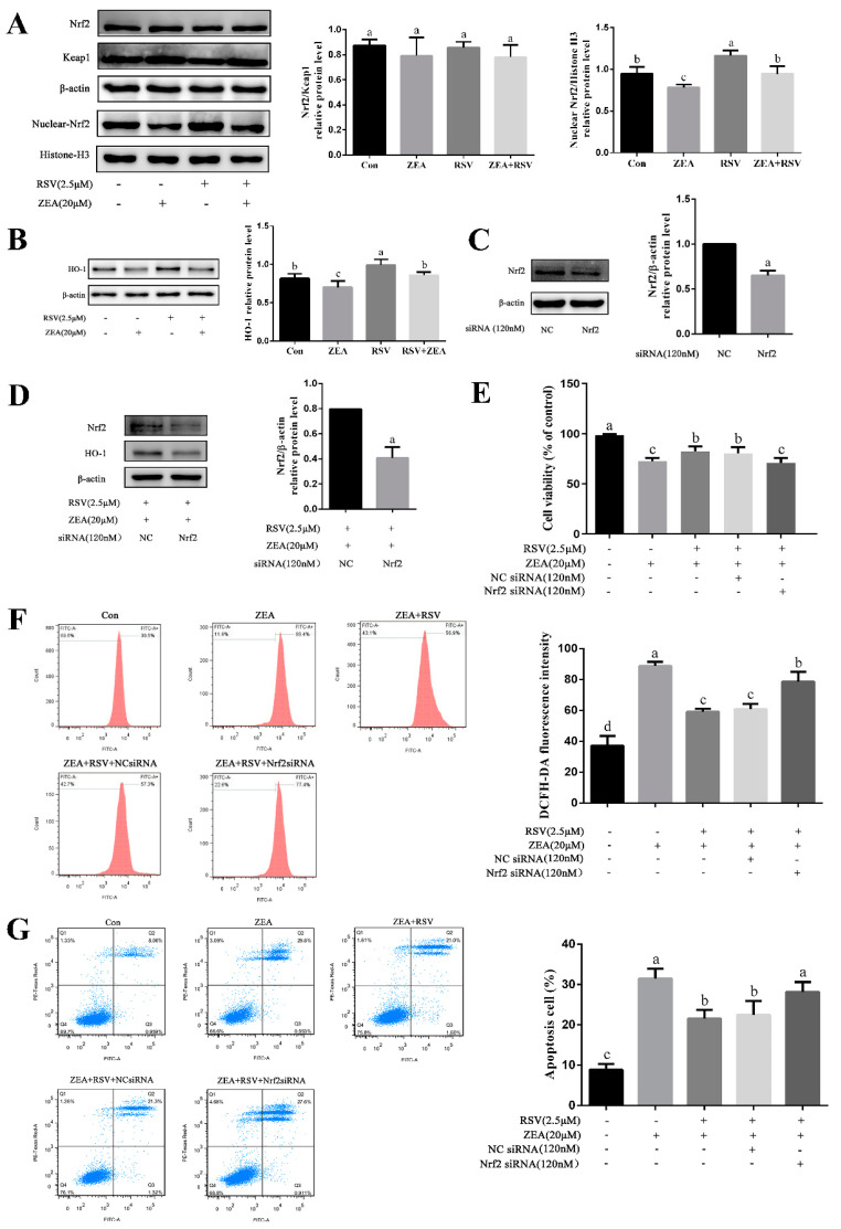
Zearalenone (ZEA) is a common mycotoxin that induces oxidative stress (OS) and affects the male reproductive system in animals. Resveratrol (RSV) has good antioxidant activity and can activate nuclear factor erythroid 2-related factor (Nrf2) to protect cells through the phosphatidylinositol 3-kinase (PI3K)/protein kinase B (Akt) signaling pathway. The objective of this study was to investigate the protective effect and the mechanism of RSV on OS and apoptosis in TM4 cells induced by ZEA. Prior to being exposed to ZEA, TM4 cells were pretreated with RSV or the PI3K/Akt inhibitor LY294002. Cell viability was measured by Cell Counting Kit-8 (CCK-8) assays. Flow cytometry was used to determine the level of apoptosis and intracellular reactive oxygen species (ROS). The expression of poly ADP-ribose polymerase (PARP), caspase-3, BCL2-associated X (Bax)/B-cell lymphoma-2 (Bcl-2), and PI3K/Akt-mediated Nrf2/heme oxygenase 1 (HO-1) signaling pathway-related proteins was evaluated by Western blotting. Nrf2 siRNA transfection and LY294002 treatment were used to investigate the role of the Nrf2/HO-1 and PI3K/Akt signaling pathways in RSV alleviation of ZEA-induced OS. The results showed that pretreatment with RSV significantly reduced the expression of apoptosis-related proteins and increased cell viability. Catalase (CAT) activity and glutathione (GSH) levels were also increased, whereas malondialdehyde (MDA) and ROS levels decreased (p < 0.05). RSV also upregulated Akt phosphorylation, Nrf2 nuclear translocation, and HO-1 expression under conditions of OS (p < 0.05). Transfection with Nrf2 siRNA abolished the protective effects of RSV against ZEA-induced cytotoxicity (p < 0.05), ROS accumulation (p < 0.05), and apoptosis (p < 0.05). LY294002 completely blocked the RSV-mediated increase in Nrf2 nuclear translocation (p < 0.05), HO-1 expression (p < 0.05), and cytoprotective activity (p < 0.05). Collectively, the above findings indicate that RSV can protect against ZEA-induced OS and apoptosis in TM4 cells by PI3K/Akt-mediated activation of the Nrf2/HO-1 signaling pathway.

Keywords: Nrf2/HO-1 signaling pathway; PI3K/Akt signaling pathway; ROS; TM4 cells; cytotoxicity; oxidative damage; resveratrol; zearalenone.


Conflict of interest statement

The authors declare no conflict of interest.

Figures

Figure 1
Figure 1
Effects of RSV on ZEA-induced TM4 cytotoxicity. TM4 cells were seeded into a 96-well plate, and the viability analysis of TM4 cells was measured by CCK-8 assay. (A) Effects of different concentrations of ZEA on TM4 cell viability. TM4 cells were treated with increasing concentrations of ZEA (0, 0.1, 1, 5, 10, 20, and 30 μmol·L−1) for 24 h. (B) Effects of different concentrations of RSV on TM4 cell viability. TM4 cells were treated with increasing concentrations of RSV (0, 1, 2.5, 5, and 7.5 μmol·L−1) for 24 h. (C) Effects of RSV on ZEA-induced TM4 cell viability. TM4 cells were exposed to ZEA (20 μmol·L−1) for 24 h after pretreatment with different concentrations of RSV for 3 h. Different letters (a, b, c, and d) represent a significant difference (p < 0.05 calculated by ANOVA for each group), while the same letters indicate no significant difference between groups.
Figure 2
Figure 2
Effects of RSV on ZEA-induced apoptosis of TM4 cells. TM4 cells were seeded into six-well plates and exposed to ZEA (20 μmol·L−1) for 24 h after pretreatment with RSV (2.5 μmol·L−1) for 3 h. (A) JC-1 staining of TM4 cells (100× magnification). TM4 cells were incubated with JC-1 (10 μg/mL) for 30 min at 37 °C and the MMP levels were analyzed by flow cytometry. A higher ratio of green/red fluorescence indicates decreased MMP. (B) Flow cytometry analysis of TM4 cell apoptosis. Cells were collected and treated with 5 μL of Annexin V-FITC and 5 μL of PI, and then incubated in the dark for 20 min at 25 °C. (C) Activity of caspase-3 in TM4 cells was measured using an active caspase-3 staining kit and detected by flow cytometry. (D) The apoptosis-related protein levels of cleaved PARP, cleaved caspase-3, and Bax/Bcl-2 in TM4 cells. Different letters (a, b, c) represent a significant difference (p < 0.05 calculated by ANOVA for each group), while the same letters indicate no significant difference between groups.
Figure 3
Figure 3
Effects of RSV on ZEA-induced oxidative stress in TM4 cells. TM4 cells were seeded into 60 mm plates and exposed to ZEA (20 μmol·L−1) for 24 h after pretreatment with RSV (2.5 μmol·L−1) for 3 h. (A) Intracellular ROS levels of TM4 cells. Cells were incubated with 10 μmol·L1 DCFH-DA for 30 min at 37 °C in the dark. The ROS levels were analyzed by flow cytometry. (B) Contents of MDA in TM4 cells. (C) Activity of CAT in TM4 cells. (D) Contents of GSH in TM4 cells. Different letters (a, b, c) represent a significant difference (p < 0.05 calculated by ANOVA for each group), while the same letters indicate no significant difference between groups.
Figure 4
Figure 4
Role of the Nrf2/HO-1 signaling pathway in alleviation of ZEA-induced TM4 cell injury by RSV. TM4 cells were exposed to ZEA (20 μmol·L−1) for 24 h after pretreatment with RSV (2.5 μmol·L−1) for 3 h after transfection with NC siRNA or Nrf2 siRNA, using Lipofectamine™ 3000 for 12 h. (A) Expression levels of Nrf2/Keap1 and nuclear-Nrf2/Histone-H3 relative proteins (total protein and nuclear protein). (B) Expression levels of HO-1 relative proteins in TM4 cells. (C,D) Expression levels of Nrf2 and HO-1 relative proteins. (E) The viability of TM4 cells detected by CCK-8 assay. (F) Intracellular ROS levels of TM4 cells were assessed using a DCFH-DA ROS assay kit. The ROS levels were analyzed by flow cytometry. (G) Flow cytometry analysis of TM4 cell apoptosis was detected using an Annexin V-FITC/PI apoptosis kit. Different letters (a, b, c, d) represent a significant difference (p < 0.05 calculated by ANOVA (A,B,EG) or a t-test (C,D) for each group), while the same letters indicate no significant difference between groups.
Figure 5
Figure 5
Role of the PI3K/Akt signaling pathway in alleviation of ZEA-induced TM4 cell injury by RSV. (A) Expression levels of p-Akt (ser 473) and Akt relative proteins in TM4 cells. TM4 cells were treated with different concentrations of RSV (0, 1, 2.5, and 5 μmol·L−1) for 24 h. (B) Expression levels of p-Akt (ser 473), p-PI3K, PI3K, and Akt relative proteins in TM4 cells. TM4 cells were pretreated with RSV (2.5 μmol·L−1) for 3 h, and then treated with or without ZEA (20 μmol·L−1) for 24 h. (C) Expression levels of p-Akt (ser 473), Akt, and Nrf2 nuclear accumulative relative proteins in TM4 cells. TM4 cells were incubated with LY294002 (10 μmol·L−1) for 1 h after pretreatment with RSV (2.5 μmol·L−1) for 3 h, then exposed to ZEA (20 μmol·L−1) for 24 h. (D) The HO-1 relative protein expression level in TM4 cells. (E) The viability of TM4 cells detected by CCK-8 assay. (F) Apoptosis of TM4 cells analyzed by flow cytometry. Cells were treated as described above and mixed with 5 μL of Annexin V-FITC and 5 μL of PI, and then incubated in the dark for 20 min at 25 °C. Different letters (a, b, c, d, e) represent a significant difference (p < 0.05 calculated by ANOVA (A,B,E,F) or a t-test (C,D) for each group), while the same letters indicate no significant difference between groups.
Figure 6
Figure 6
A proposed signaling pathway involved in RSV against ZEA-induced oxidative stress (OS) and apoptosis in TM4 cells. The schematic diagram shows that RSV induces Nrf2-mediated cytoprotective protein via activation of the PI3K/Akt signaling pathway, which protects against OS of TM4 cells. ZEA induces ROS generation results in cell apoptosis. Meanwhile, RSV activates Nrf2/HO-1 through the PI3K/Akt pathway. Activation of Nrf2/HO-1 protects against ZEA-induced apoptosis. Green arrows indicate stimulation, and red bars indicate inhibition.

Similar articles

Cited by

References

    1. Rai A., Das M., Tripathi A. Occurrence and toxicity of a fusarium mycotoxin, zearalenone. Crit. Rev. Food Sci. Nutr. 2020;60:2710–2729. doi: 10.1080/10408398.2019.1655388. - DOI - PubMed
    1. Cao L., Zhao J., Ma L., Chen J., Xu J., Rahman S.U., Feng S., Li Y., Wu J., Wang X. Lycopene attenuates zearalenone-induced oxidative damage of piglet sertoli cells through the nuclear factor erythroid-2 related factor 2 signaling pathway. Ecotoxicol. Environ. Saf. 2021;225:112737. doi: 10.1016/j.ecoenv.2021.112737. - DOI - PubMed
    1. Huang H., Lv L., Wang D., Guo B., Lv J., Luo L., Wen B., Kang Y. Biochemical and molecular responses of maize (Zea mays L.) to 1,2-dibromo-4-(1,2 dibromoethyl) cyclohexane (TBECH) diastereomers: Oxidative stress, DNA damage, antioxidant enzyme gene expression and diversity of root exudates. Sci. Total Environ. 2021;753:141872. doi: 10.1016/j.scitotenv.2020.141872. - DOI - PubMed
    1. Ren Y., Li Q., Lu L., Jin H., Tao K., Hou T. Isochamaejasmin induces toxic effects on Helicoverpa zea via DNA damage and mitochondria-associated apoptosis. Pest Manag. Sci. 2021;77:557–567. doi: 10.1002/ps.6055. - DOI - PubMed
    1. Feng N., Wang B., Cai P., Zheng W., Zou H., Gu J., Yuan Y., Liu X., Liu Z., Bian J. ZEA-induced autophagy in TM4 cells was mediated by the release of Ca2+ activates CaMKKβ-AMPK signaling pathway in the endoplasmic reticulum. Toxicol. Lett. 2020;323:1–9. doi: 10.1016/j.toxlet.2020.01.010. - DOI - PubMed

Publication types