. 2022 Nov 10;17(11):e0276694.
doi: 10.1371/journal.pone.0276694. eCollection 2022.

Long-range axonal projections of transplanted mouse embryonic stem cell-derived hypothalamic neurons into adult mouse brain

Affiliations

Long-range axonal projections of transplanted mouse embryonic stem cell-derived hypothalamic neurons into adult mouse brain

Miho Kawata et al. PLoS One. .

Abstract

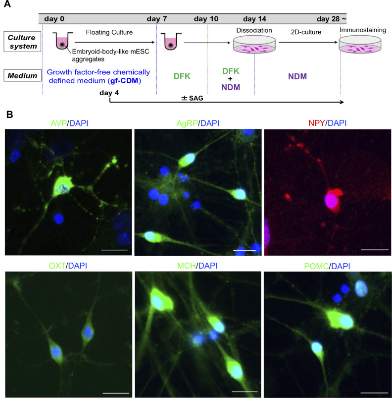
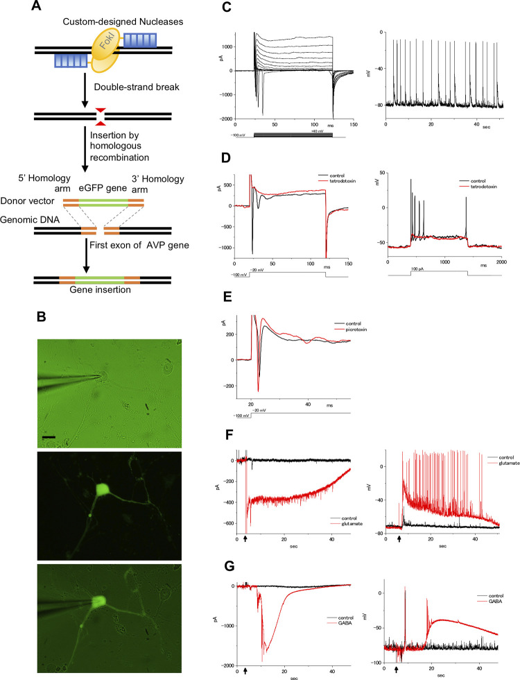
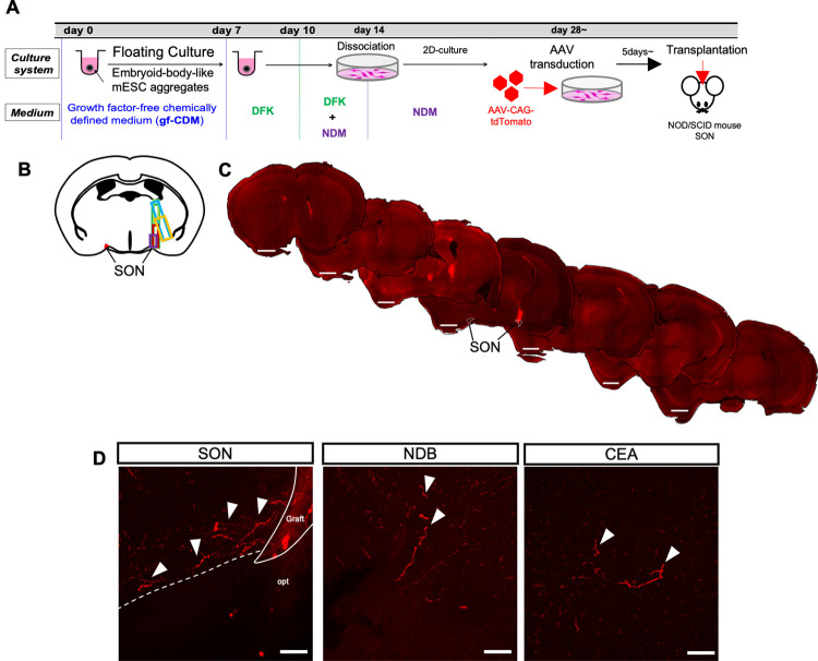
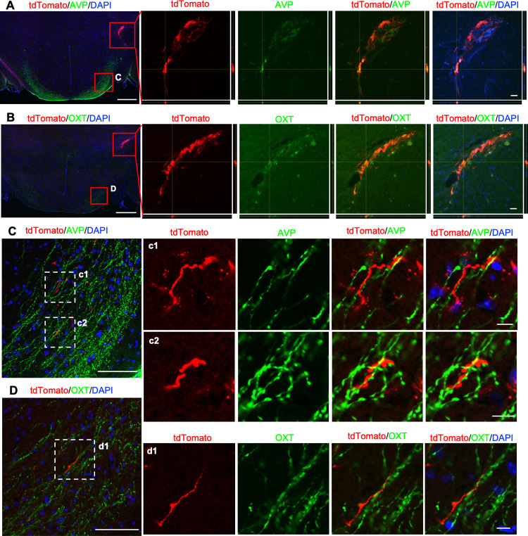
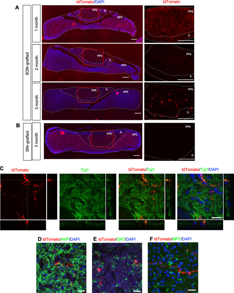
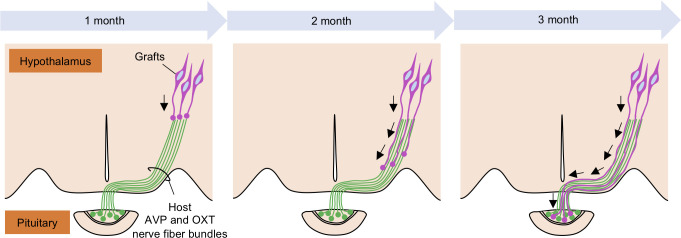
The hypothalamus is comprised of heterogenous cell populations and includes highly complex neural circuits that regulate the autonomic nerve system. Its dysfunction therefore results in severe endocrine disorders. Although recent experiments have been conducted for in vitro organogenesis of hypothalamic neurons from embryonic stem (ES) or induced pluripotent stem (iPS) cells, whether these stem cell-derived hypothalamic neurons can be useful for regenerative medicine remains unclear. We therefore performed orthotopic transplantation of mouse ES cell (mESC)-derived hypothalamic neurons into adult mouse brains. We generated electrophysiologically functional hypothalamic neurons from mESCs and transplanted them into the supraoptic nucleus of mice. Grafts extended their axons along hypothalamic nerve bundles in host brain, and some of them even projected into the posterior pituitary (PPit), which consists of distal axons of the magnocellular neurons located in hypothalamic supraoptic and paraventricular nuclei. The axonal projections to the PPit were not observed when the mESC-derived hypothalamic neurons were ectopically transplanted into the substantia nigra reticular part. These findings suggest that our stem cell-based orthotopic transplantation approach might contribute to the establishment of regenerative medicine for hypothalamic and pituitary disorders.


Conflict of interest statement

The authors have declared that no competing interests exist.

Figures

Fig 1
Fig 1. Generation of various hypothalamic neurons from mESCs.
(A) Schematic illustration of hypothalamic differentiation from mESCs. (B) Immunostaining of dissociation culture for various hypothalamic neuronal markers (AVP, AgRP, NPY, OXT, MCH and POMC). Scale bars: 20 μm.
Fig 2
Fig 2. Electrophysiological recordings of AVP GFP/+ cells.
(A) Schematic illustration of TALEN-mediated knock-in of the GFP gene into the mAVP gene locus. (B-G) Whole-cell patch-clamp recordings of the AVP GFP/+ culture cells. (B) Representative micrographs of the culture cells. The patch electrode approaches the soma of the cell from the left side (upper). Images show the GFP (middle) and Lucifer yellow fluorescence (lower) with transmitted light in the same cell as in the upper micrograph. Scale bars: 20 μm. (C) The membrane currents and spontaneous action potentials in the culture cells. Membrane currents in culture cells by depolarizing the voltage from a holding potential of –100 mV (left). Command voltages were increased in 10 mV steps from –100 mV to +40 mV. Spontaneous action potentials were observed in these cells under current-clamp conditions (right). No stimuli, such as current injections or neurotransmitters, were applied in this recording. (D) The inward current and action potential were blocked by tetrodotoxin (TTX). Membrane currents in culture cells by depolarizing the voltage from a holding potential of –100 mV (left). Command voltages were increased to −20 mV in control ringer solution (black line) or 1 μM TTX (red line). Responses to depolarization induced by injection of a 100 pA current, recorded in control ringer solution (control: black line), and the addition of 1 μM TTX (red line; right). (E) The effect of picrotoxin (PTX). Membrane currents in culture cells by depolarizing the voltage from –100 to −20 mV in control ringer solution (control: black line) or 10 μM PTX (red line). (F) The response to glutamate in the culture cells. Membrane currents were recorded under voltage-clamp conditions (left). Glutamate (up to 50 μM final concentration; red line) or ringer solution (control: black line) was applied at the arrow part. Membrane voltages were recorded under current-clamp conditions (right). (G) The response to GABA in the culture cells. Membrane currents were recorded under voltage-clamp conditions (left). GABA (up to 60 μM final concentration; red line) or ringer solution (control: black line) was applied at the arrow part. Membrane voltages were recorded under current-clamp conditions (right). Although GABA increased the membrane voltage, no action potential was generated except for immediately after the reaction (red line).
Fig 3
Fig 3. The survival and axonal projections of transplanted mESC-derived hypothalamic neurons into the SON at 3 months after transplantation.
(A) Schematic illustration of the differentiation and transplantation protocol of mESCs. (B) The map of the graft location. Each graft is shown in different-colored squares. (C) Representative overviews of grafted mESC-derived hypothalamic neurons at 3 months after transplantation. Scale bars: 1 mm. (D) Representative Z stack maximum projection images of tdTomato+ graft-derived fibers at 3 months after transplantation in the SON, NDB and CEA. opt, optic tract. White arrowheads indicate tdTomato+ fibers. Scale bars: 50 μm.
Fig 4
Fig 4. Immunohistochemistry for AVP or OXT on transplanted mESC-derived hypothalamic neurons at 3 months after transplantation.
(A, B) Left panels show immunostainings for AVP (A) or OXT (B) in the hypothalamus at 3 months after transplantation. Right panels show enlarged images of grafts depicted in the upper red squares in left panels. Z stack maximum projection images and orthogonal views show weak AVP or OXT immunoreactivities colocalized with tdTomato signals. Bottom, x-z plane; right, y-z plane. Scale bars: 500 μm (left), 20 μm (right). (C, D) Left panels show enlarged images of the supra-opticohypophysial tract depicted in lower red squares in (A) or (B). (c1, 2) and (d1) show enlarged images depicted in white dotted line squares in left panels. Scale bars: 100 μm (left), 10 μm (right).
Fig 5
Fig 5. Graft-derived axons project into the PPit.
Photographs of the pituitary at 1, 2 and 3 months after transplantation in the SON (A) or SNr (B). Left panels show the overviews of the pituitary. Right panels show enlarged images depicted in red squares in left panels. Scale bars: 200 μm. (C) Immunostaining for Tuj1 in the PPit at 3 months after transplantation. Z stack maximum projection images and orthogonal views demonstrate co-localization with the tdTomato signal and Tuj1 immunoreactivity. Bottom, x-z plane; right, y-z plane. Scale bars: 20 μm. (D, E, F) Z stack maximum projection images of the PPit immunostained for AVP (D), OXT (E) and NPY (F) at 3 months after transplantation. Scale bars: 20 μm.
Fig 6
Fig 6. Possible mechanism for the axon elongation of grafts.
A schematic illustration of the axonal elongation from grafts along the host AVP and OXT nerve fiber bundles. The nerve bundles may act as guidance cues for the grafts. It takes 3 months before the graft-derived axons reach the PPit.

Similar articles

Cited by

References

    1. Caldwell HK. Oxytocin and Vasopressin: Powerful Regulators of Social Behavior. Neuroscientist. 2017;23: 517–528. doi: 10.1177/1073858417708284 - DOI - PubMed
    1. Bonnavion P, Mickelsen LE, Fujita A, de Lecea L, Jackson AC. Hubs and spokes of the lateral hypothalamus: cell types, circuits and behaviour. J Physiol. 2016;594: 6443–6462. doi: 10.1113/JP271946 - DOI - PMC - PubMed
    1. Yeo GSH, Heisler LK. Unraveling the brain regulation of appetite: Lessons from genetics. Nat Neurosci. 2012;15: 1343–1349. doi: 10.1038/nn.3211 - DOI - PubMed
    1. Saper CB, Lowell BB. The hypothalamus. Curr Biol. 2014;24: R1111–6. doi: 10.1016/j.cub.2014.10.023 - DOI - PubMed
    1. Shapiro M, Weiss JP. Diabetes Insipidus: A Review. J Diabetes Metab. 2012;s6: 2–11. doi: 10.4172/2155-6156.S6-009 - DOI

Publication types