. 2022 Dec 23;7(78):eadf1421.
doi: 10.1126/sciimmunol.adf1421. Epub 2022 Dec 23.

SARS-CoV-2 spike conformation determines plasma neutralizing activity elicited by a wide panel of human vaccines

Affiliations

SARS-CoV-2 spike conformation determines plasma neutralizing activity elicited by a wide panel of human vaccines

John E Bowen et al. Sci Immunol. .

Abstract

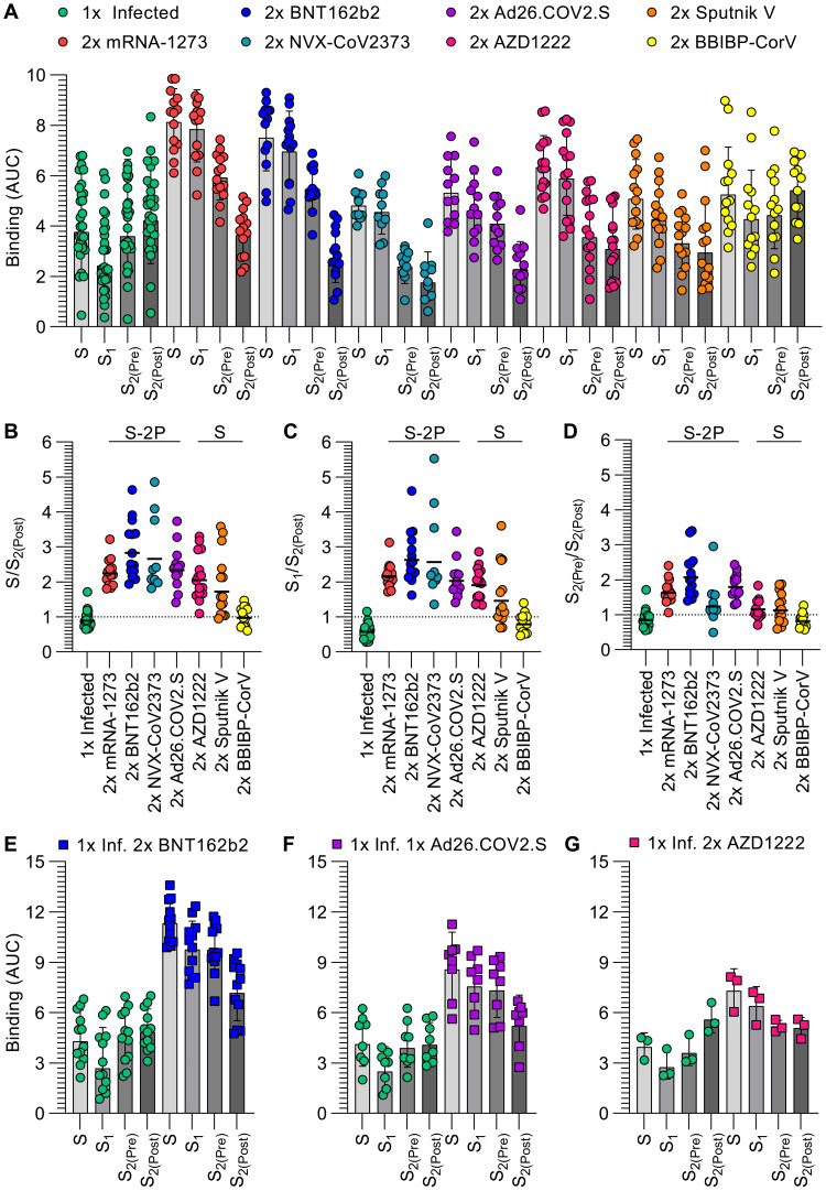
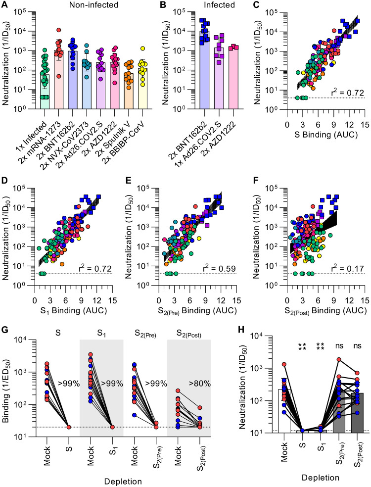
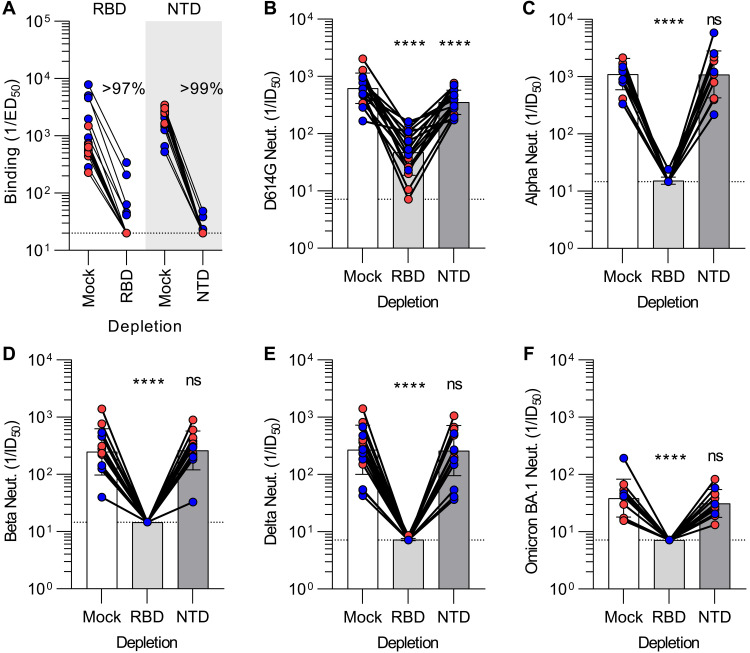
Numerous safe and effective coronavirus disease 2019 vaccines have been developed worldwide that use various delivery technologies and engineering strategies. We show here that vaccines containing prefusion-stabilizing S mutations elicit antibody responses in humans with enhanced recognition of S and the S1 subunit relative to postfusion S as compared with vaccines lacking these mutations or natural infection. Prefusion S and S1 antibody binding titers positively and equivalently correlated with neutralizing activity, and depletion of S1-directed antibodies completely abrogated plasma neutralizing activity. We show that neutralizing activity is almost entirely directed to the S1 subunit and that variant cross-neutralization is mediated solely by receptor binding domain-specific antibodies. Our data provide a quantitative framework for guiding future S engineering efforts to develop vaccines with higher resilience to the emergence of variants than current technologies.


Figures

Fig. 1.
Fig. 1.. Prefusion SARS-CoV-2 S stabilization reduces the fraction of antibodies recognizing off-target conformational states.
(A) IgG binding titers elicited by SARS-CoV-2 infection or vaccination against prefusion S (S), the S1 subunit, and the S2 subunit in the prefusion (S2(Pre)) and postfusion (S2(Post)) conformations, as measured by ELISA. Statistical analyses are shown in Tables S3-S4. (B-D) Ratio of plasma IgG binding titers against prefusion S (B), the S1 subunit (C), and the S2 subunit in the prefusion conformation (S2(Pre)) (D) over the plasma IgG binding titers against the S2 subunit in the postfusion conformation (S2(Post)). Cohorts labeled “S-2P” received vaccines encoding for or containing 2P prefusion-stabilizing S mutations whereas cohorts labeled “S” received vaccines lacking those mutations. Statistical analysis are shown in Tables S5 (E-G) IgG binding titers before and after vaccination with two doses of BNT162b2 (E), one dose of Ad26.COV2.S (F), or two doses of AZD1222 (G) in longitudinal cohorts of individuals previously infected with SARS-CoV-2. Statistical analyses are shown in Table S6 and Table S7. 1x infected samples (n = 28) were obtained 26–78 days (mean 42) post symptom onset, 2x mRNA-1273 samples (n = 14) were obtained 6–50 days (mean 15) post second dose, 2x BNT162b2 samples (n = 14) were obtained 6–33 days (mean 14) post second dose, 2x NVX-CoV2373 samples (n = 10) were obtained 17–168 days (mean 93–119) post second dose, 2x Ad26.COV2.S samples (n = 12) were obtained 12–16 days (mean 14) post second dose, 2x AZD1222 samples (n = 15) were obtained ~30 days post second dose, 2x Sputnik V samples (n = 14) were obtained 60–90 days post second dose, BBIBP-CorV samples (n = 13) were obtained 15–102 days (mean 71) post second dose, 1x Infected 2x BNT162b2 samples (n = 12) were obtained 10–32 days (mean 16) post second dose, 1x Infected 1x Ad26.COV2.S samples (n = 8) were obtained 12–112 days (mean 38) post first dose, and 1x Infected 2x AZD1222 samples (n = 3) were obtained ~30 days post second dose. Each point represents a single patient plasma sample from one representative out of at least two independent experiments consisting of different antigens, shaded bars represent the geometric mean, and error bars represent the geometric standard deviation. AUC was determined after log transforming the plasma dilution and these data are shown in Figures S5 and S6. Patient demographics are shown in Table S2.
Fig. 2.
Fig. 2.. SARS-CoV-2 neutralization is determined by S1 subunit targeting antibodies.
A-B, SARS-CoV-2 S pseudotyped VSV neutralization titers elicited by infection or vaccination (A), or vaccination following infection (B). The dotted line is the limit of detection, the colored bars are GMTs and the black error bars are geometric standard deviations. Colored points are the neutralizing geometric means of subjects after 2–4 experimental repeats consisting of different batches of pseudovirus, representative normalized curves are shown in Fig S7- S8, and statistical analyses are shown in Tables S7-S8. (C-F) Correlation between plasma neutralizing activity and prefusion S (C), S1, (D), prefusion S2 (E), and postfusion S2 (F) binding titers shown with a linear regression fit to the log of neutralization titers. The black shaded regions represent 95% confidence intervals. P < 0.0001 for all four panels. (G-H) Binding (G) and neutralization (H) titers resulting from depletion of polyclonal plasma antibodies targeting S, S1, prefusion S2, and postfusion S2. Each point is a patient plasma sample from one representative out of two independent experiments consisting of different batches of antigen and pseudovirus, shaded bars represent the geometric mean, and error bars represent the geometric standard deviation. Red points correspond to individuals vaccinated with two doses of mRNA-1273 whereas blue points correspond to individuals vaccinated with two doses of BNT162b2. Statistical significance between groups of data, relative to mock depletion, were determined by ratio paired Wilcoxon rank test and ns > 0.05, ****P < 0.0001. Mock consists of depletion carried out with beads lacking immobilized antigen. Binding data are shown in Fig S10 and dose-response neutralization curves are shown in Fig S11. Patient demographics are shown in Table S2.
Fig. 3.
Fig. 3.. Vaccine-elicited broad neutralization of SARS-CoV-2 variants is mediated by RBD-directed antibodies.
(A) Plasma IgG binding titers resulting from mock, Wuhan-Hu-1 RBD (left) and Wuhan-Hu-1 NTD (right) depletion of polyclonal antibodies. (B-F) Plasma neutralizing activity against G614 S VSV (B), Alpha S VSV (C), Beta S VSV (D), Delta S VSV (E) and Omicron BA.1 S VSV (F) after mock, Wuhan-Hu-1 RBD, or Wuhan-Hu-1 NTD depletion of polyclonal antibodies. We note that mock depleted BA.1 S VSV neutralization is dampened relative to G614 S VSV, in agreement with previous findings (53, 84, 85). Each point corresponds to a single patient plasma sample from one representative out of two independent experiments consisting of different batches of antigen and pseudovirus, shaded bars represent the geometric mean, and error bars represent the geometric standard deviation. Red points correspond to individuals vaccinated with two doses of mRNA-1273 whereas blue points correspond to individuals vaccinated with two doses of BNT162b2. Mock consists of depletion carried out with beads lacking immobilized antigen. Statistical significance between groups of data, relative to mock depletion, were determined by ratio paired Wilcoxon rank test and ns > 0.05, ****P < 0.0001. Fit binding curves are shown in Fig S12 and dose-response neutralization curves are shown in Fig S13. Patient demographics are shown in Table S2.

Update of

Similar articles

Cited by

References

    1. Walls A. C., Park Y.-J., Tortorici M. A., Wall A., McGuire A. T., Veesler D., Structure, function, and antigenicity of the SARS-CoV-2 spike glycoprotein. Cell 181, 281–292.e6 (2020). - PMC - PubMed
    1. Wrapp D., Wang N., Corbett K. S., Goldsmith J. A., Hsieh C. L., Abiona O., Graham B. S., McLellan J. S., Cryo-EM structure of the 2019-nCoV spike in the prefusion conformation. Science 367, 1260–1263 (2020). - PMC - PubMed
    1. Hoffmann M., Kleine-Weber H., Schroeder S., Krüger N., Herrler T., Erichsen S., Schiergens T. S., Herrler G., Wu N. H., Nitsche A., Müller M. A., Drosten C., Pöhlmann S., SARS-CoV-2 cell entry depends on ACE2 and TMPRSS2 and is blocked by a clinically proven protease inhibitor. Cell 181, 271–280.e8 (2020). - PMC - PubMed
    1. Hoffmann M., Kleine-Weber H., Pöhlmann S., A multibasic cleavage site in the spike protein of SARS-CoV-2 is essential for infection of human lung cells. Mol. Cell 78, 779–784.e5 (2020). - PMC - PubMed
    1. Letko M., Marzi A., Munster V., Functional assessment of cell entry and receptor usage for SARS-CoV-2 and other lineage B betacoronaviruses. Nat. Microbiol. 5, 562–569 (2020). - PMC - PubMed

Publication types