. 2022 Oct 28;9(11):601.
doi: 10.3390/vetsci9110601.

Dural Changes Induced by an Ultrasonic Bone Curette in an Excised Porcine Spinal Cord

Affiliations

Dural Changes Induced by an Ultrasonic Bone Curette in an Excised Porcine Spinal Cord

Rizou Ota et al. Vet Sci. .

Abstract

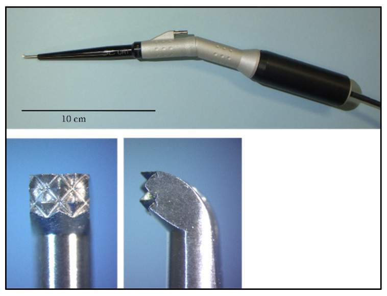
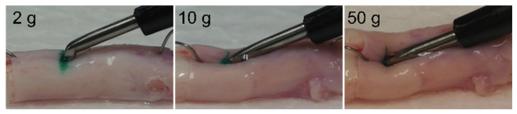
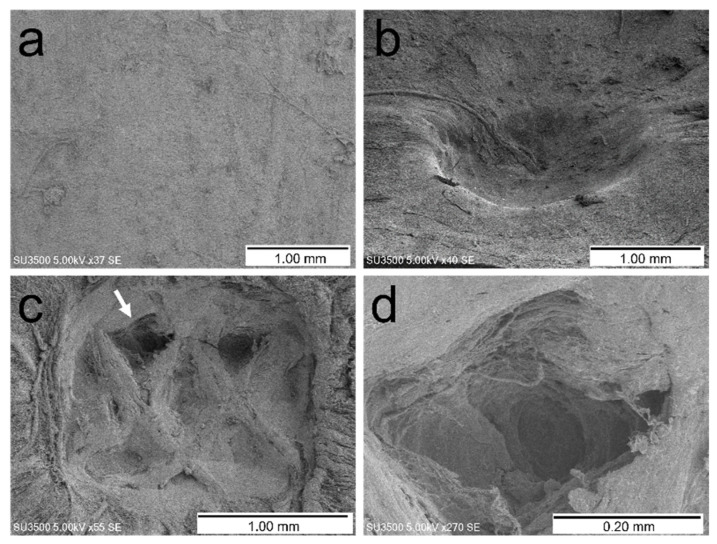
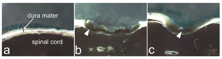
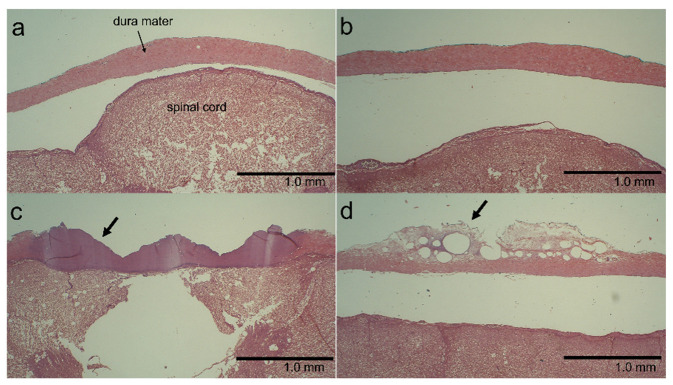
In spinal surgery, ultrasonic bone curettes are considered unlikely to cause mechanical injury to the dura; however, there is little evidence to support this claim. We investigated the effect of direct contact with an ultrasonic bone curette on the dura and the protective effect of covering the dura with a cotton pattie using an excised porcine spinal cord. The ultrasonic bone curette was pressed against the porcine spinal cord with constant force and activated for 1 s, with or without covering the dura with a cotton pattie. The dural surface and cross-section were observed using electron and light microscopy. When the ultrasonic bone curette was applied directly against the dura, most specimens showed non-perforating dural injuries. However, none of the specimens showed dural perforation. Histological changes were also observed. The use of a cotton pattie reduced the occurrence of these changes, although it did not prevent them when ultrasonic vibration was applied with a large force. We considered ultrasonic bone curettes to have a low risk of dural perforation and, thus, to be a safe surgical device as long as they did not accidentally make strong contact with the dura.

Keywords: cotton pattie; dura; spinal cord; ultrasonic bone curette.


Conflict of interest statement

The ultrasound bone curette (SonoCure®) used in this study was borrowed from Tokyo Iken Co., Ltd. (Inagi, Tokyo, Japan).

Figures

Figure 1
Figure 1
Ultrasonic bone curette handpiece (top photo) and the bone tip (bottom two photos). The width of the tip is 2 mm.
Figure 2
Figure 2
Schematic diagram of the experimental apparatus. A block of hard Styrofoam was placed on the scale, and the spinal cord specimen was fixed with needles. The ultrasonic bone curette handpiece was securely fixed to the arm of the stand, and the height of the arm was adjusted such that the ultrasonic bone curette pressed on the spinal cord specimen from above to yield a scale reading of 2, 10, or 50 g. The entire cutting surface of the handpiece tip was in contact with the spinal cord.
Figure 3
Figure 3
Examples of changes in the spinal cord when pressed by the ultrasonic bone curette at a scale reading of 2, 10, and 50 g.
Figure 4
Figure 4
Examples of changes observed in the dura mater with electron microscopy. (a) Control. (b) Dural indentation (50 g load, covered with neurosurgical cotton pattie). (c) Dural indentation or partial non-perforating wound resembling the shape of the ultrasound tip (50 g load, uncovered). (d) Partial enlargement of the site indicated by the arrow in image (c).
Figure 5
Figure 5
Examples of changes observed in the dura mater with stereomicroscopy. (a) Control. (b) Dural indentation and dark brown discoloration (arrowhead; 50 g load, no covering). (c) Dural indentation and dark brown discoloration (arrowhead; 50 g load, covered with a neurosurgical cotton pattie).
Figure 6
Figure 6
Examples of histological changes in the dura mater. (a) Control. (b) No noteworthy change is seen (10 g load, covered with a neurosurgical cotton pattie). (c) Coagulative necrosis-like change (arrow; 50 g load, uncovered). (d) Vesicle formation with edema (arrow; 2 g load, uncovered).

Similar articles

Cited by

References

    1. Moon R.D.C., Srikandarajah N., Clark S., Wilby M.J., Pigott T.D. Primary lumbar decompression using ultrasonic bone curette compared to conventional technique. Br. J. Neurosurg. 2021;35:775–779. doi: 10.1080/02688697.2020.1817321. - DOI - PubMed
    1. Petrov D., Spadola M., Berger C., Glauser G., Mahmoud A.F., O’Malley B., Malhotra N.R. Novel approach using ultrasonic bone curettage and transoral robotic surgery for en bloc resection of cervical spine chordoma: Case report. J. Neurosurg. Spine. 2019;30:788–793. doi: 10.3171/2018.11.SPINE181162. - DOI - PubMed
    1. Sun B., Xu C., Wu S., Zhang Y., Wu H., Qi M., Shen X., Yuan W., Liu Y. Efficacy and Safety of Ultrasonic Bone Curette-assisted Dome-like Laminoplasty in the Treatment of Cervical Ossification of Longitudinal Ligament. Orthop. Surg. 2021;13:161–167. doi: 10.1111/os.12858. - DOI - PMC - PubMed
    1. Nakagawa H., Uchikado H., Kim S.D., Mahadewa T., Inoue T., Mizuno J. Ultrasonic bone curettes in spinal surgery. Int. Congr. Ser. 2004;1259:445–449. doi: 10.1016/S0531-5131(03)01414-6. - DOI
    1. Rastelli M.M., Jr., Pinheiro-Neto C.D., Fernandez-Miranda J.C., Wang E.W., Snyderman C.H., Gardner P.A. Application of ultrasonic bone curette in endoscopic endonasal skull base surgery: Technical note. J. Neurol. Surg. B Skull Base. 2014;75:90–95. doi: 10.1055/s-0033-1354580. - DOI - PMC - PubMed

LinkOut - more resources