

Histological and Biochemical Analysis after Posterior Mandibular Displacement in Rats
- PMID: 36356102
- PMCID: PMC9697094
- DOI: 10.3390/vetsci9110625
Histological and Biochemical Analysis after Posterior Mandibular Displacement in Rats
Abstract
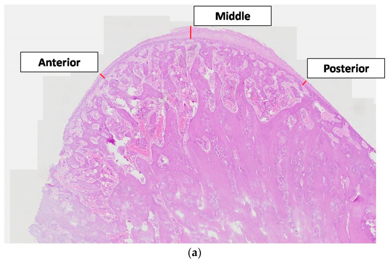
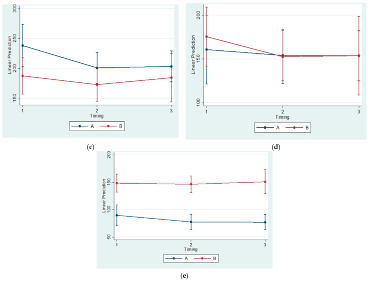
The present study aimed to investigate any biochemical and histological changes of the rat condyle and mandible in animals that had sustained mandibular growth restriction. Seventy-two male Wistar rats were divided into two equal groups, experimental and control. Each group consisted of three equal subgroups. The animals were sacrificed 30, 60, and 90 days after the start of the experiment. Blood samples were collected from the eye, and the osteoprotegerin (OPG), Receptor Activator of Nuclear Factor Kappa B Ligand (RANKL), and Macrophage Colony-Stimulating factor (MCSF)concentrations were measured by using enzyme-linked immunosorbent assay (ELISA) kits. A histological analysis was performed on the mandibular condyles. The blood serum values of OPG, RANKL, and MCSF did not exhibit any statistically significant difference between groups or subgroups. However, significant histological changes became evident after a histomorphometric condylar examination was performed. The Bone Surface/Total Surface ratio appeared reduced in the anterior and posterior regions of the condyle. In addition, the Posterior Condylar Cartilage Thickness was measured and determined to be significantly diminished. The present intervention that employed orthodontic/orthopedic devices did not prove to have any significant effect on the circulating proteins under study. Posterior displacement of the mandible may culminate only in local histological alterations in condylar cartilage thickness and its osseous microarchitecture.
Keywords: MCSF; RANKL; class III malocclusion; condylar cartilage thickness; condylar growth; mandibular posterior displacement; orthodontic treatment; osteoprotegerin; rat.
Conflict of interest statement
The authors declare no conflict of interest.
Figures


Similar articles
-
Immunohistochemical Evaluation of Bone Remodeling Following Compressive Force on Mandibular Condyle.Biology (Basel). 2025 Feb 23;14(3):228. doi: 10.3390/biology14030228. Biology (Basel). 2025. PMID: 40136485 Free PMC article.
-
[Cone-beam CT analysis of vertical control of mandible and changes of temporomandibular joint in adult patients with skeletal class Ⅱ malocclusion with high angle].Zhonghua Kou Qiang Yi Xue Za Zhi. 2022 Nov 9;57(11):1147-1155. doi: 10.3760/cma.j.cn112144-20220301-00086. Zhonghua Kou Qiang Yi Xue Za Zhi. 2022. PMID: 36379894 Chinese.
-
[Effect of gradually induced disordered occlusion on the expression of osteoprotegerin and receptor activator of nuclear factor-kappaB ligand in mandibular condylar cartilage of rats].Hua Xi Kou Qiang Yi Xue Za Zhi. 2012 Apr;30(2):119-22, 127. Hua Xi Kou Qiang Yi Xue Za Zhi. 2012. PMID: 22594224 Chinese.
-
[The effect of lowered serum levels of estrogen on the expression of ERα, ERβ, OPG and RANKL in rat mandibular condylar cartilage].Shanghai Kou Qiang Yi Xue. 2015 Aug;24(4):437-41. Shanghai Kou Qiang Yi Xue. 2015. PMID: 26383584 Chinese.
-
RANK/RANKL/OPG during orthodontic tooth movement.Orthod Craniofac Res. 2009 May;12(2):113-9. doi: 10.1111/j.1601-6343.2009.01444.x. Orthod Craniofac Res. 2009. PMID: 19419454 Review.
Cited by
-
Effects of orthodontic tooth movement on periodontal tissues after ligature-induced periodontitis through the mechanism of RANKL-induced osteoclastogenesis: an animal study.BMC Oral Health. 2025 Jul 30;25(1):1278. doi: 10.1186/s12903-025-06627-6. BMC Oral Health. 2025. PMID: 40739631 Free PMC article.
-
Growth Prediction in Orthodontics: ASystematic Review of Past Methods up to Artificial Intelligence.Children (Basel). 2025 Aug 3;12(8):1023. doi: 10.3390/children12081023. Children (Basel). 2025. PMID: 40868475 Free PMC article. Review.
-
Immunohistochemical Evaluation of Bone Remodeling Following Compressive Force on Mandibular Condyle.Biology (Basel). 2025 Feb 23;14(3):228. doi: 10.3390/biology14030228. Biology (Basel). 2025. PMID: 40136485 Free PMC article.
References
Grants and funding
LinkOut - more resources
Full Text Sources
Research Materials
