. 2022 Dec 13;55(12):2405-2418.e7.
doi: 10.1016/j.immuni.2022.10.015. Epub 2022 Nov 9.

Co-immunization with hemagglutinin stem immunogens elicits cross-group neutralizing antibodies and broad protection against influenza A viruses

Affiliations

Co-immunization with hemagglutinin stem immunogens elicits cross-group neutralizing antibodies and broad protection against influenza A viruses

Syed M Moin et al. Immunity. .

Abstract

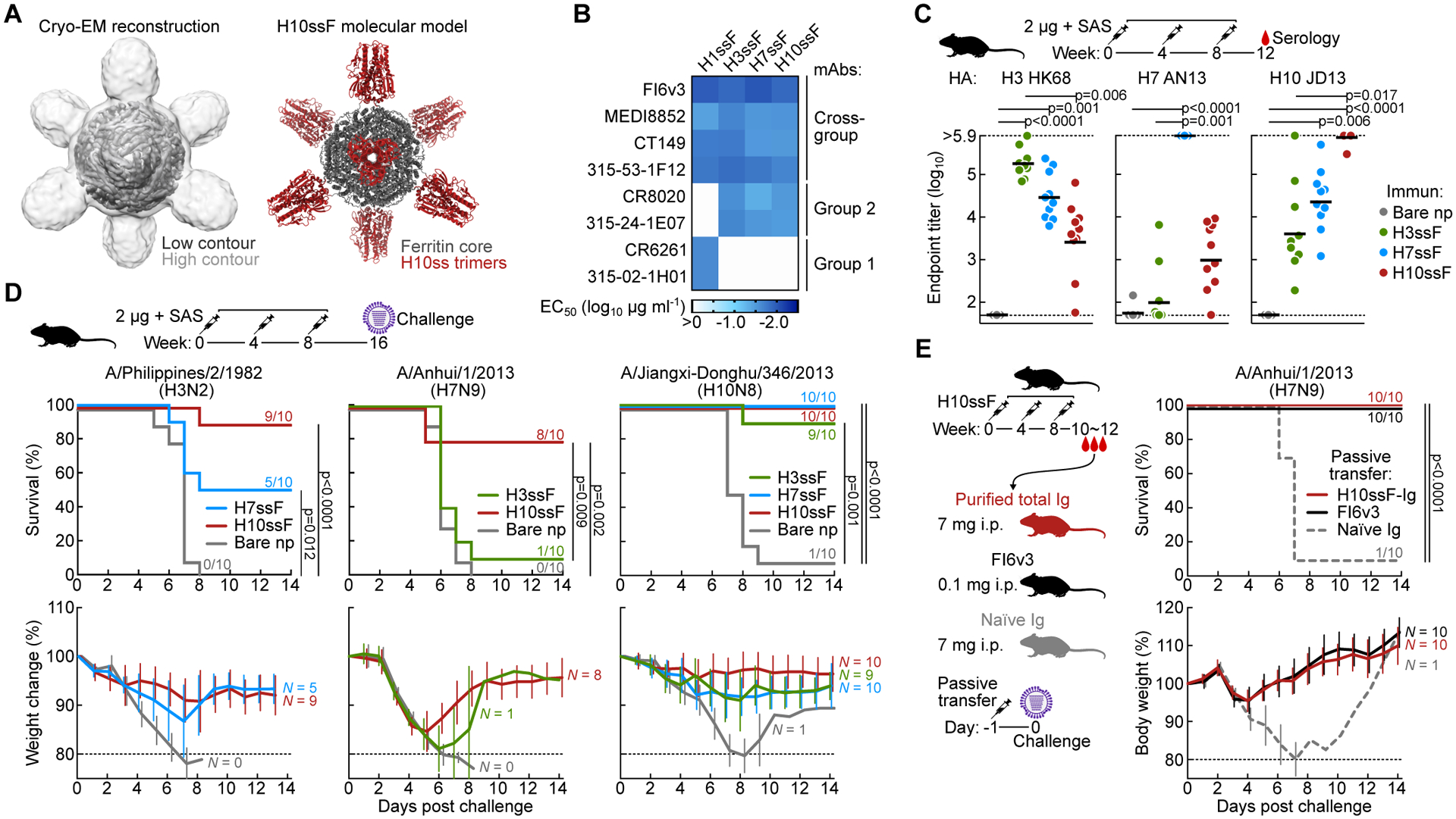
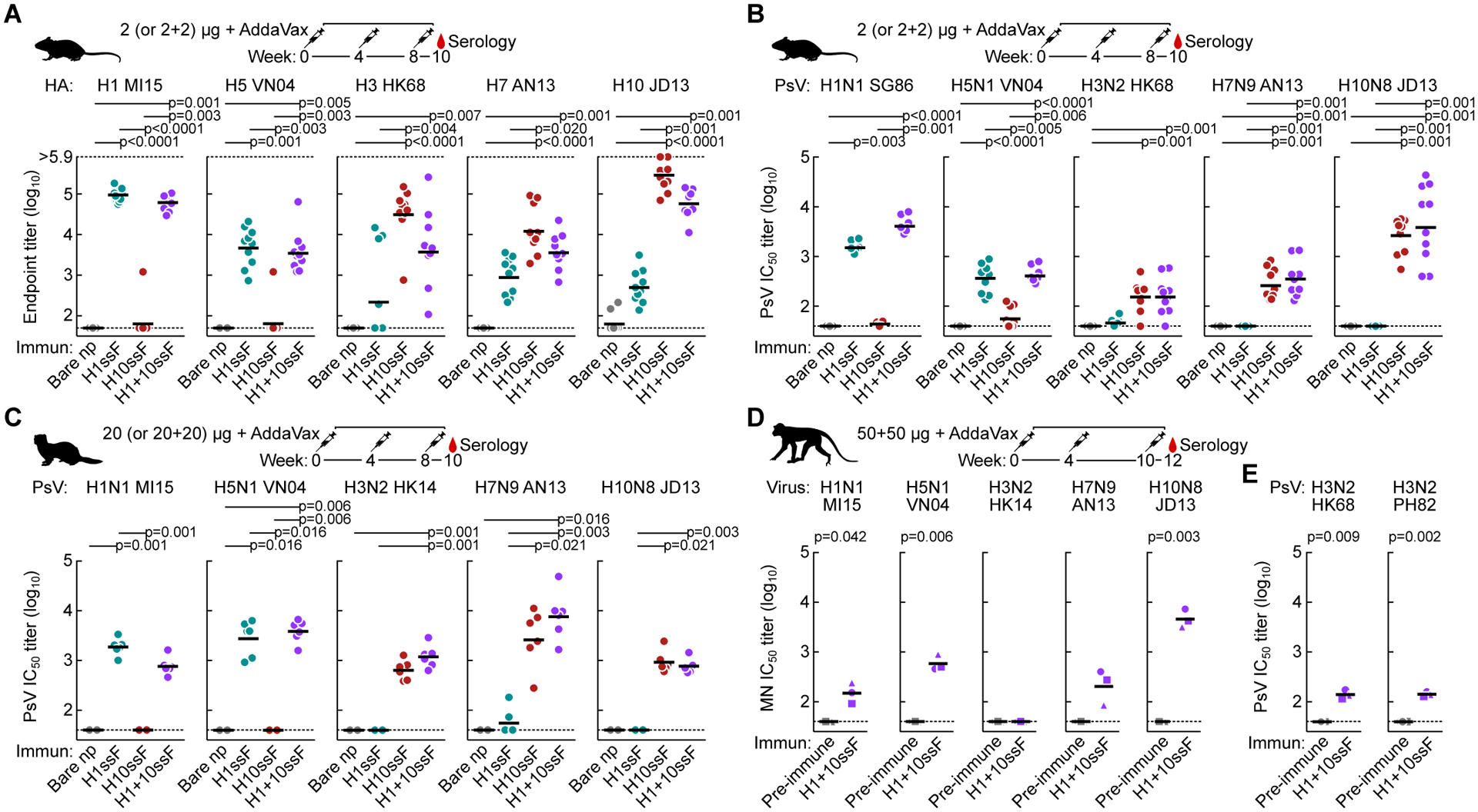
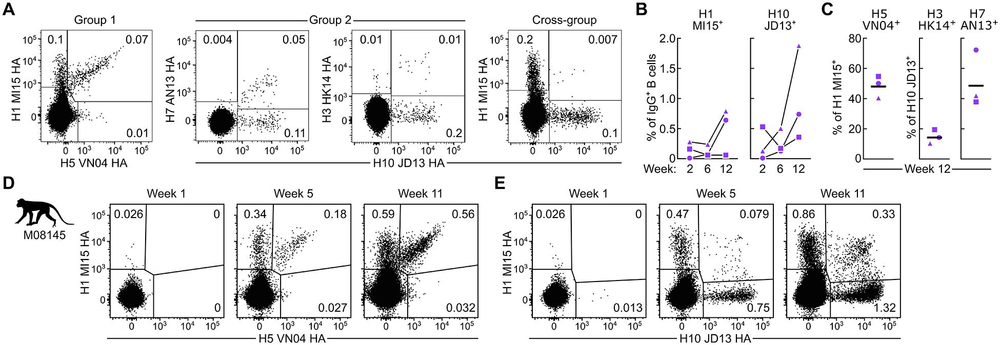
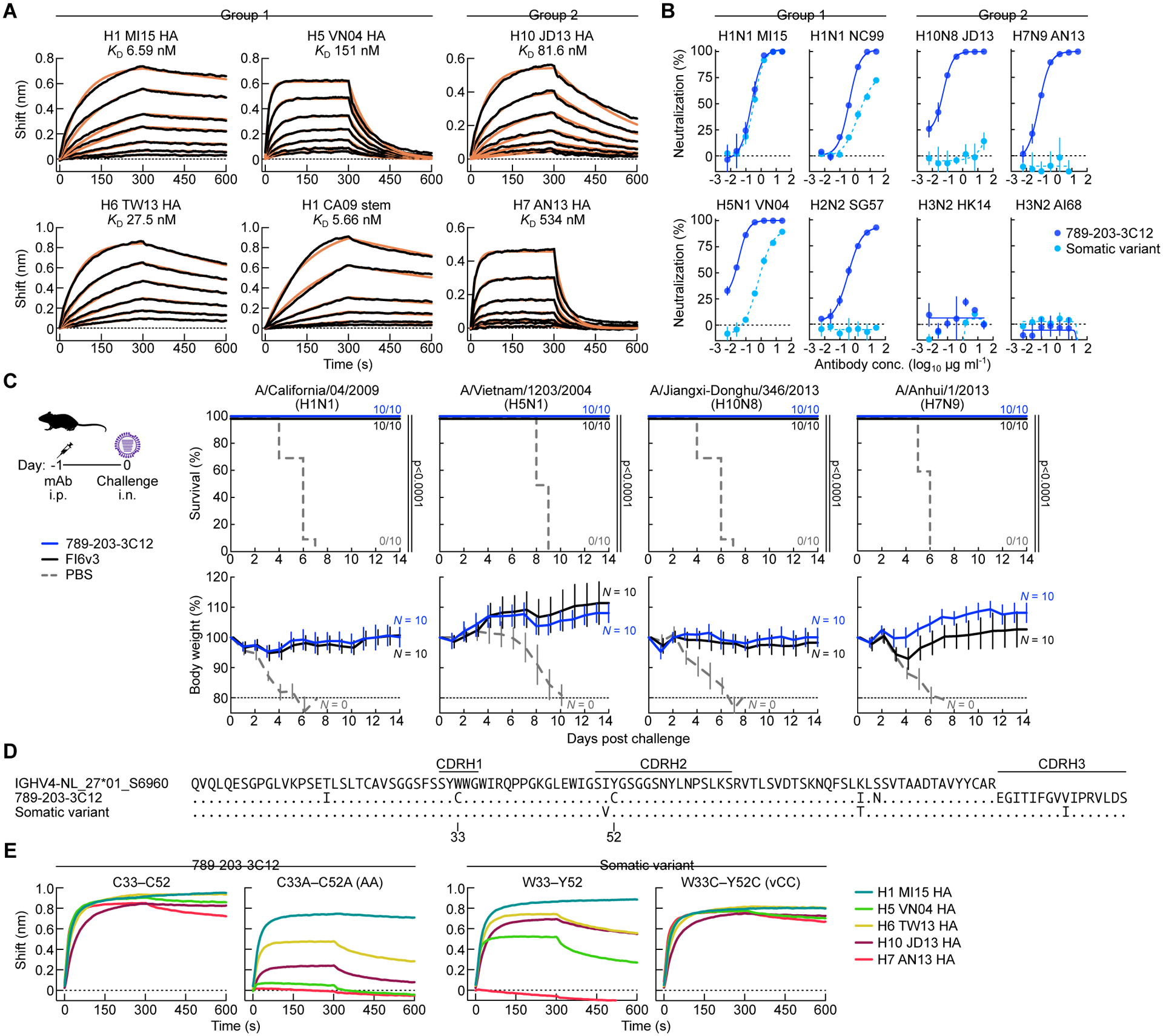
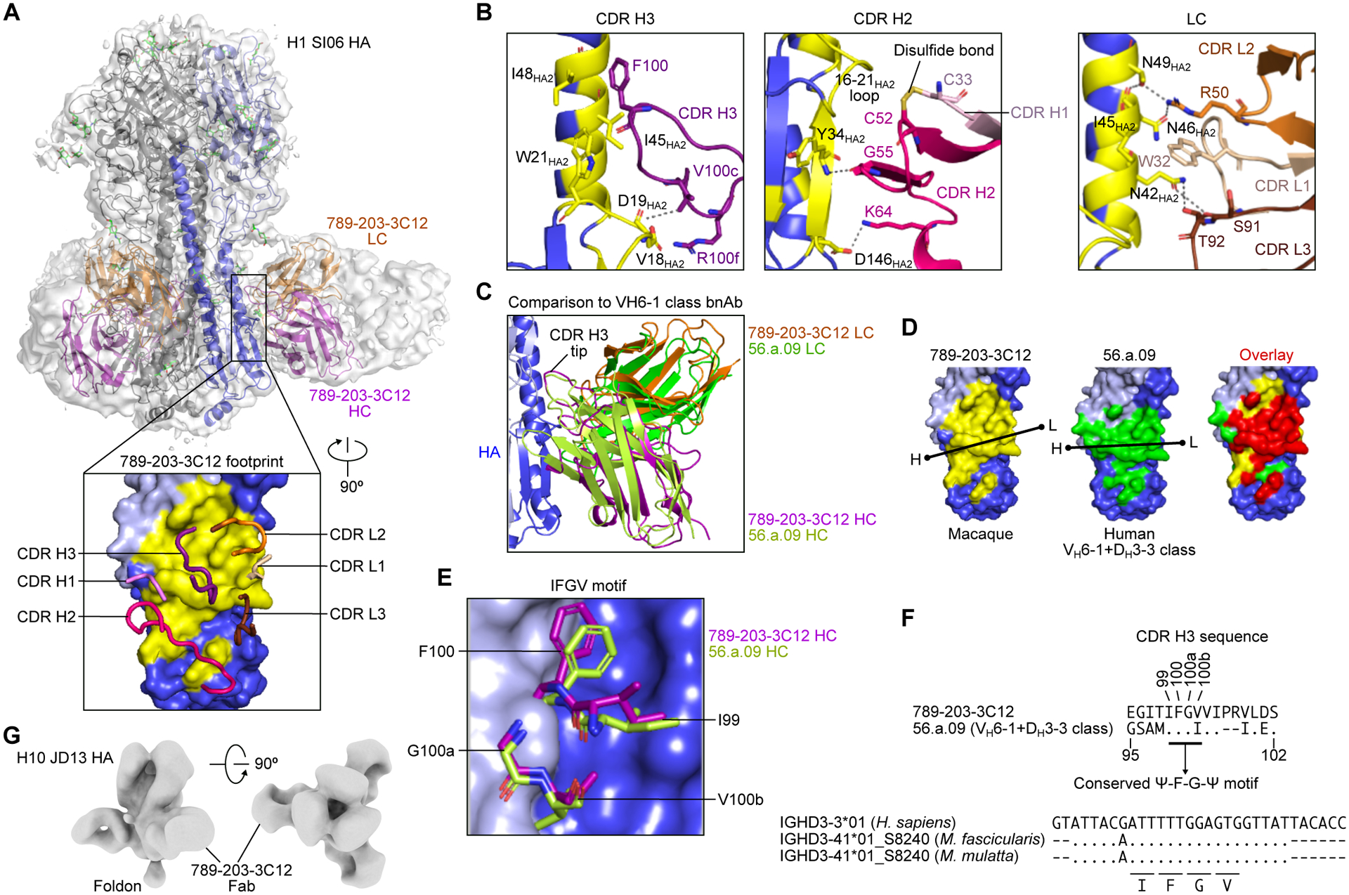
Current influenza vaccines predominantly induce immunity to the hypervariable hemagglutinin (HA) head, requiring frequent vaccine reformulation. Conversely, the immunosubdominant yet conserved HA stem harbors a supersite that is targeted by broadly neutralizing antibodies (bnAbs), representing a prime target for universal vaccines. Here, we showed that the co-immunization of two HA stem immunogens derived from group 1 and 2 influenza A viruses elicits cross-group protective immunity and neutralizing antibody responses in mice, ferrets, and nonhuman primates (NHPs). Immunized mice were protected from multiple group 1 and 2 viruses, and all animal models showed broad serum-neutralizing activity. A bnAb isolated from an immunized NHP broadly neutralized and protected against diverse viruses, including H5N1 and H7N9. Genetic and structural analyses revealed strong homology between macaque and human bnAbs, illustrating common biophysical constraints for acquiring cross-group specificity. Vaccine elicitation of stem-directed cross-group-protective immunity represents a step toward the development of broadly protective influenza vaccines.

Keywords: HA stalk; HA stem; bnAb; cryo-EM; ferritin; influenza; nanoparticle; nonhuman primate; pandemic preparedness; vaccine.


Conflict of interest statement

Declaration of interests S.M.M., J.C.B., P.D.K., J.R.M., B.S.G., and M.K. are named inventors of US patents 9,441,019, 10,137,190, 10,363,301, and 11,338,033 on influenza HA nanoparticle vaccines and stabilized HA stem trimers and of several pending applications on related technologies filed by the US Department of Health and Human Services (NIH).

Figures

Figure 1.
Figure 1.. H10-based group 2 influenza HA stem ferritin nanoparticles (H10ssF) elicits broad protective immunity against group 2 influenza A viruses.
(A) Single-particle cryo-EM structure of H10ssF. 3D reconstruction density map of H10ssF (left). The outer contour in light gray was low-pass filtered to 8 Å to reveal the HA-stem trimers, while the inner contour in dark gray indicates the 4.8 Å map of the full complex, contoured to show the ferritin core. Molecular model of the designed H10ssF showing different structural domains (right). Ferritin core and H10ss spikes are colored in gray and dark red, respectively. Cryo-EM experiment was performed once. (B) Antigenicity of HAssF determined by ELISA of HA stem-directed mAbs. mAbs FI6v3, MEDI8852, CT149, and 315–53-1F12 recognize both group 1 and group 2 HAs. mAbs CR8020 and 315–24-1E07 recognize group 2 HAs; whereas, mAbs CR6261 and 315–02-1H01 recognize group 1 HAs. Data shown are representative of three experiments. (C) Immunogenicity of group 2 HAssF in BALB/c mice. Mice (N = 10) were immunized 3 times with 2 μg H3ssF, H7ssF, or H10ssF and sera was analyzed at week 12. ELISA antibody titers to A/Hong Kong/1/1968 HA (H3 HK68), A/Anhui/1/2013 HA (H7 AN13), and A/Jiangxi-Donghu/346/13 HA (H10 JD13) are shown. Each dot represents an individual animal and the horizontal bars indicate the geometric mean of each immunization group (N = 10). Horizontal dotted lines denote higher and lower limits of detection. The mouse experiments were independently performed at least three times, and representative data are shown. Statistical analysis was performed by Kruskal-Wallis test with Dunn’s multiple comparison post-hoc test. (D) Protective efficacy of group 2 HAssF in mice. BALB/c mice immunized with H3ssF, H7ssF, or H10ssF (N = 10) were experimentally infected intranasally with either A/Philippines/2/1982 (H3N2), A/Anhui/1/2013 (H7N9), or A/Jiangxi-Donghu/346/2013 (H10N8) viruses 8 weeks post the last immunization. Data shown are representative of at least two independent experiments. (E) Passive transfer experiment of purified hyperimmune Igs followed by experimental H7N9 infection. Igs (7 mg) of H10ssF-immunized or naïve mice were passively given to naïve BALB/c mice (N = 10) intraperitoneally 24 h prior to infecting with A/Anhui/1/2013 (H7N9). mAb FI6v3 (5 mg kg−1) was used as positive control. The passive transfer experiment was performed once. Multiple comparisons of Kaplan-Meier curves were performed by the log-rank test with Bonferroni correction (D and E). See also Figures S1, S2, and S3.
Figure 2.
Figure 2.. Co-immunization of group 1 and group 2 HAssF elicits broadly cross-reactive and neutralizing antibody responses in mice, ferrets, and NHPs.
(A) Cross-reactive HA-binding antibody responses to multiple group 1 and group 2 HAs. Sera (N = 10) were collected after 3 immunizations with H1ssF, H10ssF, H1+10ssF, or bare nanoparticles. ELISA antibody responses were assessed for A/Michigan/45/2015 (H1 MI15), A/Vietnam/1203/2004 (H5 VN04), H3 HK68, H7 AN13, and H10 JD13 HA.Mouse experiments were performed at least twice, and representative data are shown. All assays were repeated twice, and representative data are shown. (B and C) Neutralizing antibody responses to pseudotyped lentiviruses expressing multiple group 1 and group 2 HAs in mice (B) and ferrets (N = 6) (C). Pseudotype neutralization assays were performed by using HA and NA from A/Singapore/1/1986 (H1N1 SG86), H1N1 MI15, H5N1 VN04, H3N2 HK68, H7N9 AN13, and H10N8 JD13. Mouse experiments were performed at least twice, and representative data are shown. Ferret experiment was performed once. All assays were repeated twice, and representative data are shown. (D and E) Neutralizing antibody responses in NHPs. Cynomolgus macaques (N = 3) were immunized with H1+10ssF. Serum neutralizing activity as measured by the reporter-based microneutralization assay (D)(Creanga et al., 2021) or Pseudotype neutralization assay (E). H1N1 MI15, H5N1 VN04, A/Hong Kong/4801/2014 (H3N2 HK14), H7N9 AN13, and H10N8 JD13 reporter influenza viruses were used for microneutralization assay. Pseudotype viruses were made using HA and NA from H3N2 HK68 and A/Philippines/2/1982 (H3N2 PH82) (E). Different symbols are used to identify individual animals. Black horizontal lines indicate geometric mean titers for each respective group. Horizontal dotted lines denote higher and lower limits of detection (A) or lower limit of detection (B to D). Statistical analysis was performed by Kruskal-Wallis test with Dunn’s multiple comparison post-hoc test (A to C) or by paired t-test (D). NHP experiment was performed once. All assays were repeated twice and representative data are shown. See also Figure S3.
Figure 3.
Figure 3.. Co-immunization with group 1 and group 2 HAssF induces cross-reactive HA-specific B cells in NHPs.
(A) Detection of HA-specific B cells by flow cytometry. PBMCs from NHPs (N = 3) at two weeks after the second immunization were analyzed by using fluorescently-labeled HA trimer probes. (B) Frequencies of homologous H1+ and H10+ B cells in PBMCs. Percentage of H1+ or H10+ among IgG+ B cells are shown. (C) Frequencies of cross-reactive B cells. Percentage of H5 positivity among H1+ B cells (left), and percentages of H7+ or H3+ among H10+ B cells (right) are shown. Horizontal lines indicate the geometric mean of a group. (D,E) Kinetics of cross-reactive B cell responses in macaque M08145. PBMCs taken at one week after each immunization were analyzed by flow cytometry with H1 MI15 and H5 VN04 (D), or H1 MI15 and H10 JD13 (E) HA trimer probes. Flow cytometry experiments were performed twice, and representative data are shown. See also Figure S4.
Figure 4.
Figure 4.. The NHP mAb 789–203-3C12 shows broad neutralization breadth, potency, and protective efficacy against group 1 and group 2 influenza A viruses.
(A) HA-binding kinetics of Fab 789–203-3C12 determined by BLI. Recombinant HA of multiple group 1 and group 2 subtypes or the stabilized HA stem were immobilized on biosensors. Experimental datasets were fitted with the binding equations describing a 1:1 interaction to obtain apparent affinities. Experiments were performed twice, and representative data are shown. (B) Neutralization profile of mAb 789–203-3C12 and its somatic variant antibody. Neutralization curves were generated by the reporter-based microneutralization assay(Creanga et al., 2021). Experiments were performed twice, and representative data are shown. (C) Protective efficacy of mAb 789–203-3C12 in mice. 789–203-3C12 or control FI6v3 antibodies were passively administered (10 mg kg−1) to recipient BALB/c mice (N = 10) intraperitoneally 24 h prior to infecting with multiple group 1 and group 2 subtype viruses. Multiple comparisons of Kaplan-Meier curves were performed by the log-rank test with Bonferroni correction. Mouse experiment was performed once. (D) Sequence alignment of the germline-encoded IGHV4-NL_27*01_S6960 heavy chain, 789–203-3C12, and its somatic variant. Dots denote identical residues to the IGHV4-NL_27*01_S6960 whereas the residues that are different from the IGHV4-NL_27*01_S6960 are indicated. CDR loop regions are shown by horizontal lines (Kabat). (E) Binding profile of mAb 789–203-3C12 and the somatic variant antibody with their CDR H1 and CDR H2 variants. HA-binding was measured using BLI. Recombinant HAs were immobilized on biosensors. All the mAbs used in the assay were tested as IgG. Experiments were performed twice, and representative data are shown.
Figure 5.
Figure 5.. Structural characterization of NHP mAb 789–203-3C12 identifies conserved DH motifs between human and NHP bnAbs for HA recognition.
(A) Overall structure of the 789–203-3C12 Fab bound to the H1 SI06 HA trimer. The HA trimer is shown in gray, with the protomer with bound antibody colored blue. HA1, HA2, and antibody heavy and light chains are depicted as ribbons colored light blue, blue, purple, and orange, respectively. A semitransparent surface representation of the entire complex is also shown in gray. A 90° rotated view of the interface with interacting CDR loops shown as worms and HA as surface representation (inset). The epitope of 789–203-3C12 is colored in yellow. Cryo-EM experiment was performed once. (B) Detailed interactions of 789–203-3C12 CDR loops with HA. The formation of a disulfide bond between CDR H1 and H2 loops are shown in yellow (middle panel). Hydrogen bonds and salt bridges are indicated as dashed lines. (C) Comparison of the binding mode of 789–203-3C12 antibody with the human VH6–1+DH3–3 class bnAb human 56.a.09 on HA. (D) The footprints for 789–203-3C12 (yellow) and 56.a.09 (green) are mapped onto the HA surface. A black line indicates the orientation of the antibody defined by the line connecting Cα atoms of heavy chain Cys22 (dot with letter H) and light chain Cys23 (dot with letter L). The overlapping epitope was colored in red (right). (E) Structural arrangement of the “Ψ-F-G-Ψ” motif in the 789–203-3C12 and 56.a.09. Ψ is I, V, L, or M. (F) Sequence comparison of the CDR H3 of 789–203-3C12 and 56.a.09 (top) and sequence alignment of human IGHD3–3*01 and IGHD3–41*01_S8240 genes from macaque species (bottom). Amino acid positions shown are based on the Kabat numbering. Dots denote identical nucleotides to the human IGHD3–3*01 whereas the nucleotides that are different from the human IGHD3–3*01 are indicated. Codons encoding the IFGV motif are underlined. (G) Negative stain EM 3D reconstruction of 789–203-3C12 Fab bound to the H10 JD13 HA trimer. EM experiment was performed once. See also Figure S5 and Tables S1, S2, and S3.

Similar articles

Cited by

References

    1. Adams PD, Gopal K, Grosse-Kunstleve RW, Hung L-W, Ioerger TR, McCoy AJ, Moriarty NW, Pai RK, Read RJ, Romo TD, et al. (2004). Recent developments in the PHENIX software for automated crystallographic structure determination. J. Synchrotron Radiat 11, 53–55. 10.1107/s0909049503024130. - DOI - PubMed
    1. Andrews SF, Joyce MG, Chambers MJ, Gillespie RA, Kanekiyo M, Leung K, Yang ES, Tsybovsky Y, Wheatley AK, Crank MC, et al. (2017). Preferential induction of cross-group influenza A hemagglutinin stem-specific memory B cells after H7N9 immunization in humans. Sci Immunol 2, eaan2676. 10.1126/sciimmunol.aan2676. - DOI - PubMed
    1. Avnir Y, Watson CT, Glanville J, Peterson EC, Tallarico AS, Bennett AS, Qin K, Fu Y, Huang C-Y, Beigel JH, et al. (2016). IGHV1–69 polymorphism modulates anti-influenza antibody repertoires, correlates with IGHV utilization shifts and varies by ethnicity. Sci. Rep 6, 20842. 10.1038/srep20842. - DOI - PMC - PubMed
    1. Barad BA, Echols N, Wang RY-R, Cheng Y, DiMaio F, Adams PD, and Fraser JS (2015). EMRinger: side chain–directed model and map validation for 3D cryo-electron microscopy. Nat. Methods 12, 943–946. 10.1038/nmeth.3541. - DOI - PMC - PubMed
    1. Bernstein DI, Guptill J, Naficy A, Nachbagauer R, Berlanda-Scorza F, Feser J, Wilson PC, Solórzano A, Van der Wielen M, Walter EB, et al. (2020). Immunogenicity of chimeric haemagglutinin-based, universal influenza virus vaccine candidates: interim results of a randomised, placebo-controlled, phase 1 clinical trial. Lancet Infect. Dis 20, 80–91. 10.1016/S1473-3099(19)30393-7. - DOI - PMC - PubMed

Publication types

Substances