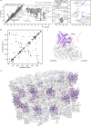
Magic-angle-spinning NMR structure of the kinesin-1 motor domain assembled with microtubules reveals the elusive neck linker orientation
- PMID: 36357375
- PMCID: PMC9649657
- DOI: 10.1038/s41467-022-34026-w
Magic-angle-spinning NMR structure of the kinesin-1 motor domain assembled with microtubules reveals the elusive neck linker orientation
Abstract
Microtubules (MTs) and their associated proteins play essential roles in maintaining cell structure, organelle transport, cell motility, and cell division. Two motors, kinesin and cytoplasmic dynein link the MT network to transported cargos using ATP for force generation. Here, we report an all-atom NMR structure of nucleotide-free kinesin-1 motor domain (apo-KIF5B) in complex with paclitaxel-stabilized microtubules using magic-angle-spinning (MAS) NMR spectroscopy. The structure reveals the position and orientation of the functionally important neck linker and how ADP induces structural and dynamic changes that ensue in the neck linker. These results demonstrate that the neck linker is in the undocked conformation and oriented in the direction opposite to the KIF5B movement. Chemical shift perturbations and intensity changes indicate that a significant portion of ADP-KIF5B is in the neck linker docked state. This study also highlights the unique capability of MAS NMR to provide atomic-level information on dynamic regions of biological assemblies.
© 2022. The Author(s).
Conflict of interest statement
The authors declare no competing interests.
Figures


Similar articles
-
Comprehensive structural model of the mechanochemical cycle of a mitotic motor highlights molecular adaptations in the kinesin family.Proc Natl Acad Sci U S A. 2014 Feb 4;111(5):1837-42. doi: 10.1073/pnas.1319848111. Epub 2014 Jan 21. Proc Natl Acad Sci U S A. 2014. PMID: 24449904 Free PMC article.
-
The structure of apo-kinesin bound to tubulin links the nucleotide cycle to movement.Nat Commun. 2014 Nov 14;5:5364. doi: 10.1038/ncomms6364. Nat Commun. 2014. PMID: 25395082
-
Mapping the structural and dynamical features of kinesin motor domains.PLoS Comput Biol. 2013;9(11):e1003329. doi: 10.1371/journal.pcbi.1003329. Epub 2013 Nov 7. PLoS Comput Biol. 2013. PMID: 24244137 Free PMC article.
-
Overview of the mechanism of cytoskeletal motors based on structure.Biophys Rev. 2018 Apr;10(2):571-581. doi: 10.1007/s12551-017-0368-1. Epub 2017 Dec 12. Biophys Rev. 2018. PMID: 29235081 Free PMC article. Review.
-
Why are ATP-driven microtubule minus-end directed motors critical to plants? An overview of plant multifunctional kinesins.Funct Plant Biol. 2020 May;47(6):524-536. doi: 10.1071/FP19177. Funct Plant Biol. 2020. PMID: 32336322 Review.
Cited by
-
Distinct Clinical Phenotypes in KIF1A-Associated Neurological Disorders Result from Different Amino Acid Substitutions at the Same Residue in KIF1A.Biomolecules. 2025 May 2;15(5):656. doi: 10.3390/biom15050656. Biomolecules. 2025. PMID: 40427549 Free PMC article.
-
A structural and dynamic visualization of the interaction between MAP7 and microtubules.Nat Commun. 2024 Mar 2;15(1):1948. doi: 10.1038/s41467-024-46260-5. Nat Commun. 2024. PMID: 38431715 Free PMC article.
-
Structural basis of protein condensation on microtubules underlying branching microtubule nucleation.Nat Commun. 2023 Jun 21;14(1):3682. doi: 10.1038/s41467-023-39176-z. Nat Commun. 2023. PMID: 37344496 Free PMC article.
-
Microtubule association induces a Mg-free apo-like ADP pre-release conformation in kinesin-1 that is unaffected by its autoinhibitory tail.Nat Commun. 2025 Jul 5;16(1):6214. doi: 10.1038/s41467-025-61498-3. Nat Commun. 2025. PMID: 40617823 Free PMC article.
-
Dipolar Recoupling in Rotating Solids.Chem Rev. 2024 Nov 27;124(22):12844-12917. doi: 10.1021/acs.chemrev.4c00373. Epub 2024 Nov 6. Chem Rev. 2024. PMID: 39504237 Free PMC article. Review.
References
-
- Vale RD. The molecular motor toolbox for intracellular transport. Cell. 2003;112:467–480. - PubMed
-
- Wittmann T, Hyman A, Desai A. The spindle: a dynamic assembly of microtubules and motors. Nat. Cell Biol. 2001;3:E28–E34. - PubMed
-
- Howard J, Hyman AA. Dynamics and mechanics of the microtubule plus end. Nature. 2003;422:753–758. - PubMed
-
- Hirokawa N, Noda Y, Tanaka Y, Niwa S. Kinesin superfamily motor proteins and intracellular transport. Nat. Rev. Mol. Cell Biol. 2009;10:682–696. - PubMed
Publication types
MeSH terms
Substances
Grants and funding
LinkOut - more resources
Full Text Sources
Miscellaneous

