

Radiographical characteristics and traction duration of impacted maxillary canine requiring surgical exposure and orthodontic traction: a cross-sectional study
- PMID: 36357464
- PMCID: PMC9649639
- DOI: 10.1038/s41598-022-23232-7
Radiographical characteristics and traction duration of impacted maxillary canine requiring surgical exposure and orthodontic traction: a cross-sectional study
Abstract
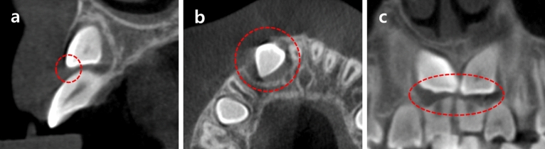
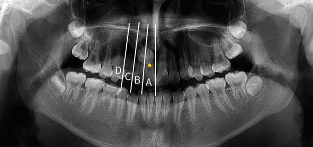
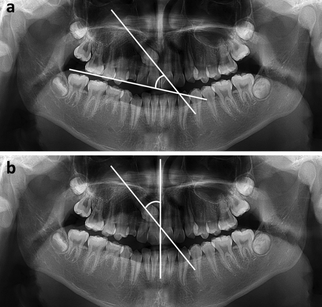
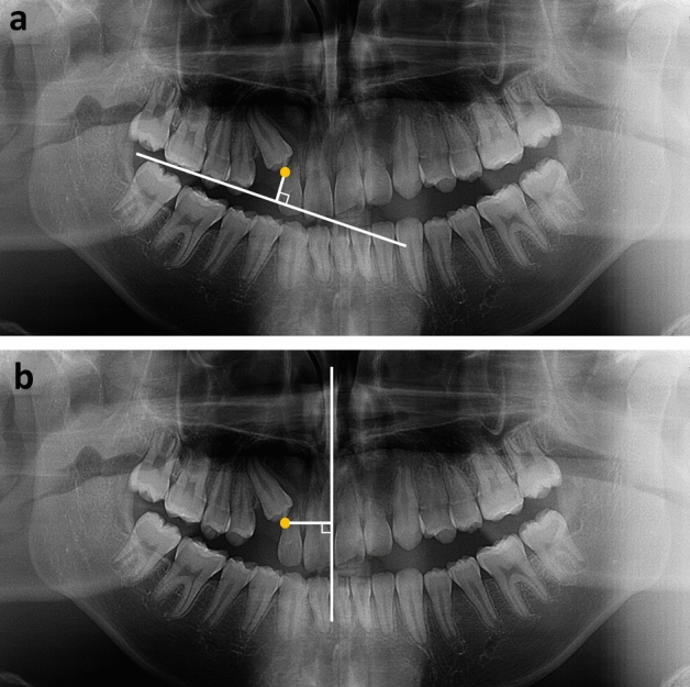
This cross-sectional study aimed to classify the radiographical characteristics of impacted maxillary canines that were surgically exposed following orthodontic traction and to find out which factor is most closely related to traction duration. This study enrolled 74 patients with 87 maxillary canines. Cone-beam computed tomography images, panoramic radiographs, and medical records were analyzed. Cystic-appearing lesion and resorption of adjacent roots were observed in 26.4% and 23.0% of cases, respectively. Impacted maxillary canines were mostly distributed in the lateral incisor area. The mean (± standard deviation) traction duration for the 47 teeth that met the study criteria was 13.9 (± 8.9) months. Impacted maxillary canines treated with surgical exposure and orthodontic traction showed increasing possibilities of palatal impaction and resorption of the adjacent root as they were located mesially (p < 0.05). The distance from the occlusal plane to the impacted maxillary canine showed the strongest positive correlation with traction duration (r = 0.519, p < 0.01). When establishing treatment plans for patients with impacted maxillary canines, distance from the occlusal plane to the impacted canines, rather than the angle, should be considered in predicting the duration of treatment.
© 2022. The Author(s).
Conflict of interest statement
The authors declare no competing interests.
Figures
References
-
- Hamada Y, Timothius CJC, Shin D, John V. Canine impaction—A review of the prevalence, etiology, diagnosis and treatment. Semin. Orthod. 2019;25(2):117–123. doi: 10.1053/j.sodo.2019.05.002. - DOI
-
- Arboleda-Ariza N, Schilling J, Arriola-Guillén LE, Ruíz-Mora GA, Rodríguez-Cárdenas YA, Aliaga-Del Castillo A. Maxillary transverse dimensions in subjects with and without impacted canines: A comparative cone-beam computed tomography study. Am. J. Orthod. Dentofacial Orthop. 2018;154(4):495–503. doi: 10.1016/j.ajodo.2017.12.017. - DOI - PubMed
Publication types
MeSH terms
LinkOut - more resources
Full Text Sources
Miscellaneous
