. 2022 Nov 10;12(1):19185.
doi: 10.1038/s41598-022-23689-6.

A new catalytic site functioning in antigen cleavage by H34 catalytic antibody light chain

Affiliations

A new catalytic site functioning in antigen cleavage by H34 catalytic antibody light chain

Emi Hifumi et al. Sci Rep. .

Abstract

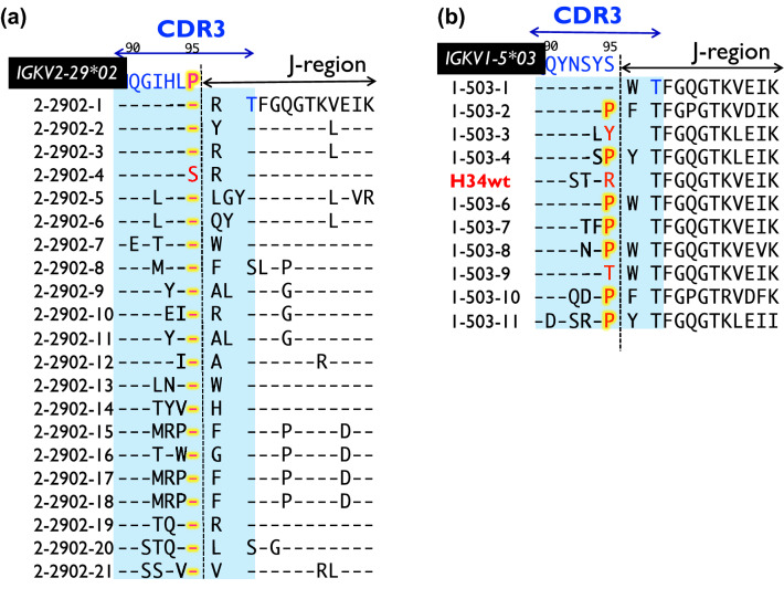
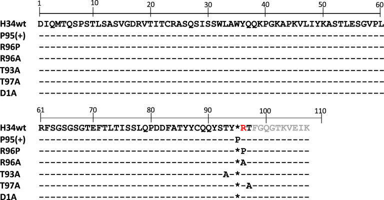
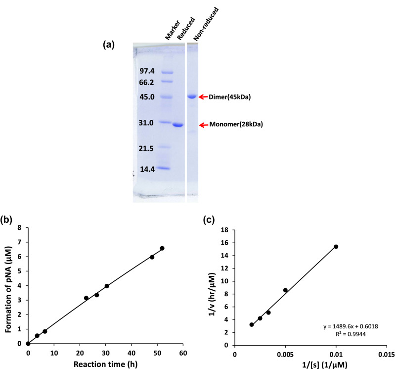
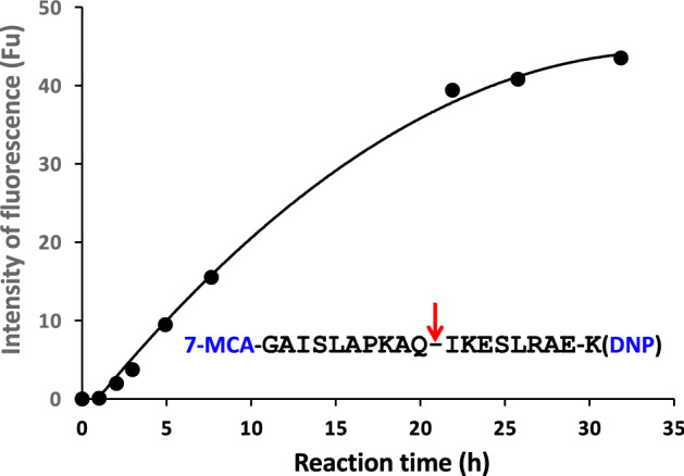
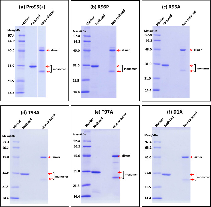
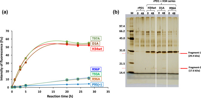
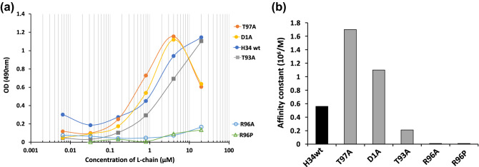
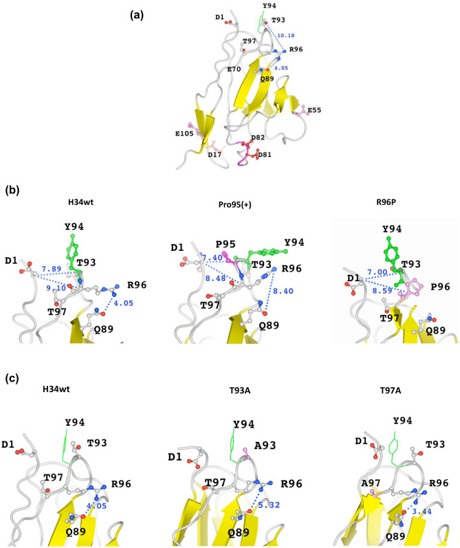
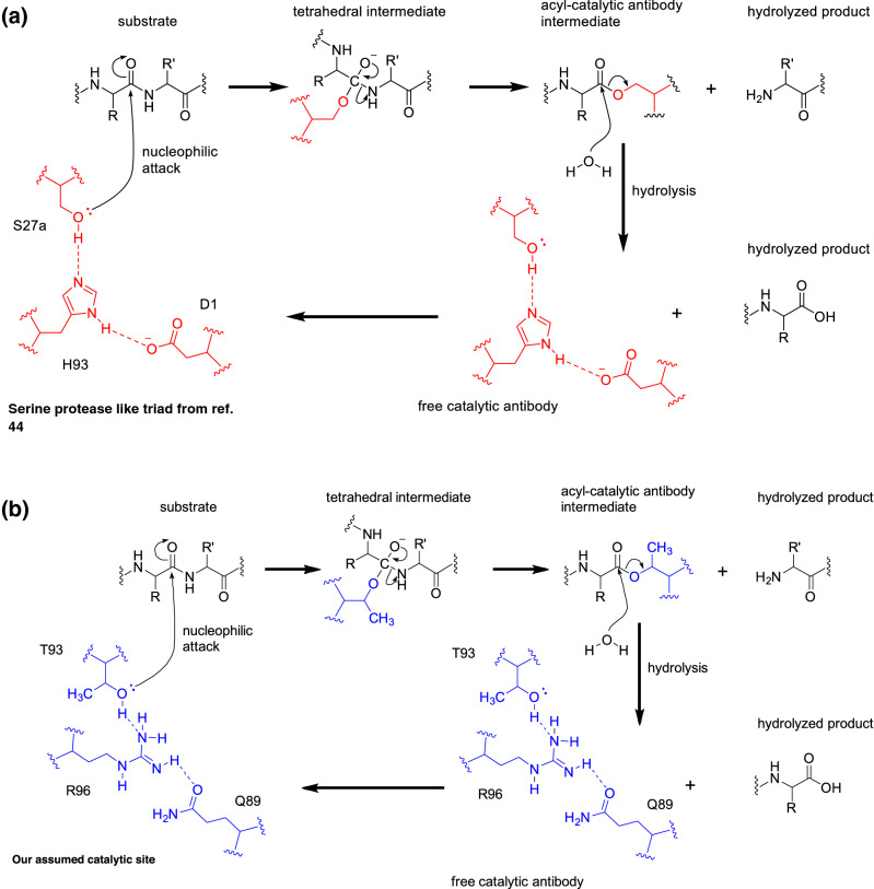
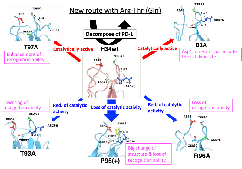
The cleavage reactions of catalytic antibodies are mediated by a serine protease mechanism involving a catalytic triad composed of His, Ser, and Asp residues, which reside in the variable region. Recently, we discovered a catalytic antibody, H34 wild type (H34wt), that is capable of enzymatically cleaving an immune-check point PD-1 peptide and recombinant PD-1; however, H34wt does not contain His residues in the variable region. To clarify the reason behind the catalytic features of H34wt and the amino acid residues involved in the catalytic reaction, we performed site-directed mutagenesis focusing on the amino acid residues involved in the cleavage reaction, followed by catalytic activity tests, immunological reactivity evaluation, and molecular modeling. The results revealed that the cleavage reaction by H34wt proceeds through the action of a new catalytic site composed of Arg, Thr, and Gln. This new scheme differs from that of the serine protease mechanism of catalytic antibodies.


Conflict of interest statement

The authors declare no competing interests.

Figures

Figure 1
Figure 1
Comparison of amino acid sequences of CDR-3 among germline genes. (a) IGKV2-29*02: the amino acid sequences of germline gene IGKV2-29*02 and 21 human light chain clones. 20 out of 21 clones possess Pro95 residue at the 95th position, indicating that Pro95 is highly conserved. (b) IGKV1-5*03: IGKV1-5*03 does not contain Pro95; a Ser residue is placed at this position. Although the residue is replaced by somatic mutation, the Ser is replaced with Pro in 64% of the clones (seven out of eleven clones). Antibody light chains tend to contain Pro at the 95th position in CDR-3.
Figure 2
Figure 2
Amino acid sequences for H34wt and the mutants. The amino acid sequences of the variable region of H34wt and the mutants. There are no histidine residues in the variable region. Another feature of H34wt is the lack of Pro95, which is present in many antibodies. The amino acid residues considered to participate in the catalytic reaction are mutated to Pro or Ala. DNA sequences for H34wt and the mutants are deposited in DDBJ/GenBank/EMBL (see Data availability statement).
Figure 3
Figure 3
(a) Results of SDS-PAGE analysis for H34wt: SDS-PAGE analysis (12% gel) for H34wt after a size-exclusion chromatography is shown. A band of dimer at approximately 45 kDa is observed under the non-reduced condition. Under the reduced condition, only a single band of approximately 28 kDa is detected, which corresponds to the monomer. Bands other than the monomer of the light chains are essentially absent under either reduced or non-reduced condition. (b) Time course of the cleavage of the synthetic substrate Arg-pNA by H34wt. Reaction conditions: H34wt: 10 μM. Arg‐pNA: 200 μM. Reaction temperature: 37 °C. Reaction volume: 200 µL. Arg(R)‐pNA (trypsin‐like substrate) was used as the substrate in 50 mM/Tris-100 mM/Glycine-Tween-20 buffer (TGT) buffer. The reaction was performed in triplicate in a 96-well microplate. The figure shows that the cleavage reaction proceeds almost linearly depending on the reaction time. (c) Kinetics. Lineweaver–Burk plot for cleavage of Arg-pNA by H34wt. H34wt: 10uM. Arg-pNA: 100–800 µM. Reaction temperature: 37 °C. The plot presents a linear relationship, indicating that the cleavage reaction is enzymatic and in accordance with the Michaelis–Menten mechanism. Km = 2.48 × 10−3 (M) and kcat = 2.7 × 10−3 (/min).
Figure 4
Figure 4
The time course for the cleavage of FRET-PD1 by H34wt: the cleavage reaction was performed with 5 µM H34wt using 25 µM of FRET-PD1 (PD-1; aa 123-140). The reaction was performed in duplicate. H34wt cleaves the FRET-PD1 peptide in a linear fashion up to 20 h of the reaction time, following which it reaches a plateau.
Figure 5
Figure 5
SDS-PAGE for mutants. SDS-PAGE (12% gel) with CBB staining was carried out following the procedure described in Fig. 3a. Each mutant was purified by cation chromatography. A band of dimer at approximately 45 kDa is observed under non-reduced condition. (a) Pro95(+); A faint band at ~ 50 kDa is observed over the dimer band at 45 kDa under non-reduced condition. Under the reduced condition, a band is observed at approximately 29 kDa; no other bands are detected. (b) R96P; In this sample, a faint band at ~ 50 kDa is observed over the dimer band at 45 kDa under non-reduced condition. Under the reduced condition, a band is observed at approximately 28 kDa; no other bands are detected. (c) R96A; Under both reduced and non-reduced conditions, no other bands except R96A mutant are observed. (d) T93A; Under both reduced and non-reduced conditions, no other bands except R96A mutant are observed. (e) T97A; A faint band at ~ 40 kDa below the dimer band (44 kDa) is detected under the non-reduced condition, whereas the monomer band is clearer compared with that of other mutants. A faint band caused by a small amount of impurities is observed at ~ 16 kDa. (f) D1A; under both reduced and non-reduced conditions, no other bands except R96A mutant are observed.
Figure 6
Figure 6
(a) Time course of the cleavage reaction for FRET-PD1 peptide by mutants. FRET-PD1: 25 µM. H34wt & mutants: 5 µM. In this experiment, H34wt was tested together with the mutants for ensuring accurate comparison among the samples. H34wt and the mutants of D1A and T97A show high catalytic activity. In contrast, three mutants of R95P, R95A, and T93A exhibit low catalytic activity. The catalytic activity of P95(+) is substantially suppressed. (b) Cleavage of recombinant PD-1 molecule. Recombinant PD-1 (rPD-1): 1 µM. H34wt, D1A, R96A and T97A: 0.5 µM. Reaction temperature: 37 °C. The SDS-PAGE (12% gel) experiment was performed under reduced condition, and the visualization was performed by silver staining. The cleavage reaction by H34wt and the several mutants were carried out over 48 h of incubation. The robust bands at approximately 29-kDa represent the monomer band of H34wt and/or the mutants. In H34wt light chain, a band at 17.5 kDa, which corresponds to the fragment of rPD1 (see ref.), is detected at 48 h of incubation; however, it is not detected at 0 h of incubation. Similar results are observed for D1A mutant, where the same band at 17.5 kDa is detected. In contrast, no band at 17.5 kDa is detected in the case of the mutant R96A, indicating that R96A does not cleave rPD-1. These results are consistent with the cleavage activity for FRET-PD1. Both H34wt and D1A, but not R96A, cleaves FRET-PD1 peptide with high activity.
Figure 7
Figure 7
(a) ELISA: Coated antigen (rPD1): 2 µg/mL and 50 µL/well. Concentration of H34wt and the mutants: 0.0064–20 µM. Second antibody: POD-goat IGG to human Kappa chain (1/2000 dilution). H34wt shows the typical sigmoid curve of ELISA. Both T97A and D1A mutants shows the sigmoid curve with prozone phenomena. The mutants are divided into two groups. The first group including H34wt, T97A, D1A, and T93A shows binding affinity to rPD-1, while the second group including R96A and R96P hardly binds to rPD-1. This means that Arg96 is strongly involved in the recognition of rPD-1. (b) Comparison of affinity constants. The affinity constants, which were calculated from the results of (a), are presented for comprehensive comparison. It is clear that the replacement of The93 and Arg96 to Ala (or Pro) decreased the antigen recognition.
Figure 8
Figure 8
Molecular modeling. Numerical number is the distance between two atoms (Å). (a) H34wt: Asp17, Asp81, Asp82, Glu55, Glu70, and Glu105 residues capable of pulling a proton of Arg96 are present in the variable region; however, they are too far to interact with Arg96. The carbonyl of Gln89 residue can interact with the hydrogen of amino group of Arg96. The distance between them is 4.05 Å, which is preferable for interaction. (b) Comparison of H34wt, Pro95(+), and R96P: the conformation of Tyr94 residue of H34wt and Pro95(+) is considerably changed from the vertical position in H34wt to the horizontal position in Pro95(+). Additionally, the conformation of Arg96 is considerably altered. The conformational changes between H34wt and R96P are small, except the replacement of the residue. In H34wt, Pro(95+) and R96P, the distances of Cα between D1-T93 and D1-R96 are in the range within 1.0 Å, suggesting small changes in the clones. (c) Comparison of H34wt, T93A, and T97A: the conformations of Arg96 are slightly different among the three light chains; however, those of Tyr94 are not considerably changed. The distance between Q89(O)-R96(N) becomes longest (5.32 Å) in T93A among three clones. The β-sheet content appears to increase in the mutants T93A and T97A compared with that in H34wt.
Figure 9
Figure 9
Linear Free Energy Relationship (LFER). LFER exists among the H34wt, T97A, D1A, and T93A clones, suggesting that features of these four clones are slightly different. In contrast, T93A and R96P are out of the linearity, indicating that the features of these two light chains must be hugely different from the above four clones.
Figure 10
Figure 10
Reaction scheme by serine protease-like triad and catalytic site of H34wt. (a) In the case of serine protease-like triad, the hydroxyl group of the catalytic Ser of the catalytic antibody directly initially attacks the cleavable bond. (b) In the case of H34wt, similarly, the hydroxyl group of the catalytic Thr of H34wt directly attacks the scissile bond. Then, finally, the deacylation of the acyl catalytic antibody intermediate with water forms a second product and regenerates the free catalytic antibody.
Figure 11
Figure 11
Interrelations of features of H34wt and the mutants. Mutagenesis in this study clarified the amino acid residues constructing the catalytic site, which is composed of Arg96, Thr93, and Gln89, revealing a new catalytic site for the cleavage of PD-1, which is different from the serine protease triad. The figure shows the interrelation among H34wt and the mutants; in addition to structural changes, several crucial changes are detected in terms of chemical and immunological characters. The number indicates the distance between the Arg and Thr residues. The figure shows the interrelation among H34wt and the mutants; in addition to structural changes, several crucial changes are detected in terms of chemical and immunological characters. The number indicates the distance between the Arg and Thr residues.

Similar articles

Cited by

References

    1. Paul S, et al. Catalytic hydrolysis of vasoactive intestinal peptide by human autoantibody. Science. 1989;244:1158–1162. - PubMed
    1. Hifumi E, Kondo H, Mitsuda Y, Uda T. Catalytic features of monoclonal antibody i41SL1-2 subunits. Biotechnol. Bioeng. 2003;84:485–493. - PubMed
    1. Mei S, Mody B, Eklund SH, Paul S. Vasoactive intestinal peptide hydrolysis by antibody light chains (Communication) J. Biol. Chem. 1991;266:15571–15574. - PubMed
    1. Gao QS, et al. Molecular cloning of a proteolytic antibody light chain. J. Biol. Chem. 1994;269:32389–32393. - PubMed
    1. Taguchi H, et al. Autoantibody-catalyzed hydrolysis of Amyloid β peptide. J. Biol. Chem. 2008;283:4714–4722. - PubMed

Publication types

MeSH terms