

Case Reports
doi: 10.1038/s41380-022-01853-8.
Epub 2022 Nov 10.
Obsessive-compulsive symptoms in two patients with strategic basal ganglia lesions
Affiliations
- PMID: 36357672
- PMCID: PMC9908536
- DOI: 10.1038/s41380-022-01853-8
Item in Clipboard
Case Reports
Obsessive-compulsive symptoms in two patients with strategic basal ganglia lesions
Mol Psychiatry.
2023 Feb.
No abstract available
Conflict of interest statement
RD: Lecture fees from Roche, Alexion, Bayer, and Sanofi. Travel grant from Biogen. KD: Steering Committee Neurosciences, Janssen. Speaker fees from Janssen. VAC: Consultation work for CereGate (Munich, Germany), Cortec (Freiburg, Germany), and Inbrain (Barcelona, Spain). Honoraria for Talks (Boston Scientific, USA). IITs in DBS with Medtronic (USA) and Boston Scientific (USA). All other authors declare no potential competing interests.
Figures
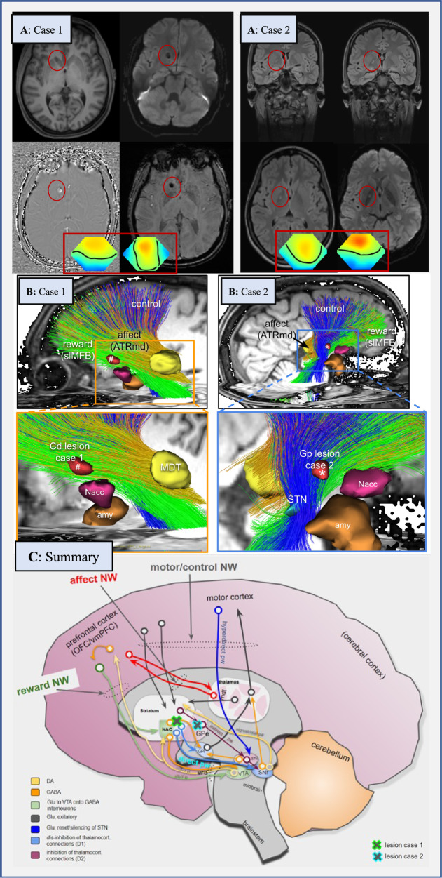
A: Case 1 shows a lesion in the right caudate nucleus with a size of 8.9 × 5 mm. The lesion was classified as a probable microbleeding, but calcification as a late consequence of microbleeding could not be definitively ruled out. Case 2 presents with a FLAIR-hyperintense lesion in the globus pallidus on the right (defect with a gliotic rim in the direction of a lenticulostriate vessel). Because Case 2 had febrile seizures during her first decade, and since then chronic obsessive-compulsive disorder (OCD), a post-inflammatory origin could be plausible. Framed below in red are the topographies of the prominent theta electroencephalography (EEG) components for the two patients. In both cases the pronounced frontocentral theta activity is suggestive of sleepiness. Topographies are against common average reference with standard orientation (top = front), positive excursions in red, negative in blue. Zero potential is indicated by a black line. Spontaneous EEG was recorded during a 12-minute series of standard maneuvers (eyes open, eyes closed, and hyperventilation) according to the 10–20 system (21 head channels), leading to 21 independent component analysis (ICA) components. Artifact-free sections across the whole recording length were submitted to ICA training. ICA maps as well as associated EEG time courses were reviewed and components classified by their topography, reaction to the maneuvers, and spectrum. Additionally, intermittent rhythmic delta/theta activity was detected in the artifact-free sections using an automated algorithm. The software used for these analyses is a combination of the EEG data processor “avg_q” (https://github.com/berndf/avg_q ) and custom python scripts. B: Case 1 has tractographic rendition of streamlines involved in strategic unilateral lesions in the inferior and anterior caudate nucleus (nucleus accumbens spared) (#, right side). Tractographic analysis of Case 2 shows a pallidal lesion (*) involving the “motor-control network” (blue fibers). C: Interplay of three involved networks (“affect, control, and reward network”) within the whole “OCD framework” (existing of the “affect, control, reward, and default-mode network” [2, 13]) at distinct network hubs (taken and altered from [13]). The locations of lesions in Cases 1 and 2 are shown as green/blue crosses. Abbreviations: Amy amygdala, ATR anterior thalamic radiation, ATRmd anterior thalamic radiation from dorsomedial thalamus, BNST bed nucleus of stria terminalis, Cd caudate nucleus, DA dopamine, EEG electroencephalography, GABA gamma-aminobutyric acid, GPe globus pallidus externus, GPi globus pallidus internus, Glu glutamate, imMFB infero-medial MFB, ICA independent component analysis, MDT mediodorsal thalamus, MFB medial forebrain bundle, NAC/Nacc nucleus accumbens, NW network, OCD obsessive-compulsive disorder, OFC orbitofrontal cortex, pw pathway, slMFB superolateral branch of the medial forebrain bundleMFB, SNr substantia nigra, STN subthalamic nucleus, VTA ventral tegmental area, vmPFC ventromedial prefrontal cortex.
Comment on
-
Toward a neurocircuit-based taxonomy to guide treatment of obsessive-compulsive disorder.Mol Psychiatry. 2021 Sep;26(9):4583-4604. doi: 10.1038/s41380-020-01007-8. Epub 2021 Jan 7. Mol Psychiatry. 2021. PMID: 33414496 Free PMC article. Review.
References
-
- Coenen VA, Schlaepfer TE, Sajonz B, Döbrössy M, Kaller CP, Urbach H, et al. Tractographic description of major subcortical projection pathways passing the anterior limb of the internal capsule. Corticopetal organization of networks relevant for psychiatric disorders. Neuroimage Clin. 2020;25:102165. doi: 10.1016/j.nicl.2020.102165. - DOI - PMC - PubMed
-
- Saraiva LC, Sato JR, Cappi C. Probing the genetic and molecular correlates of connectome alterations in obsessive-compulsive disorder. Mol Psychiatry. 2022. 10.1038/s41380-022-01590-y. - PubMed
Publication types
MeSH terms
LinkOut - more resources
Full Text Sources
Medical
