Review
. 2023 Apr;20(4):263-277.
doi: 10.1038/s41569-022-00796-5. Epub 2022 Nov 10.

Temporary mechanical circulatory support devices: practical considerations for all stakeholders

Affiliations
Review

Temporary mechanical circulatory support devices: practical considerations for all stakeholders

Benjamin S Salter et al. Nat Rev Cardiol. 2023 Apr.

Abstract

Originally intended for life-saving salvage therapy, the use of temporary mechanical circulatory support (MCS) devices has become increasingly widespread in a variety of clinical settings in the contemporary era. Their use as a short-term, prophylactic support vehicle has expanded to include procedures in the catheterization laboratory, electrophysiology suite, operating room and intensive care unit. Accordingly, MCS device design and technology continue to develop at a rapid pace. In this Review, we describe the functionality, indications, management and complications associated with temporary MCS, together with scenario-specific utilization, goal-directed development and bioengineering of future devices. We address various considerations for the use of temporary MCS devices in both prophylactic and rescue scenarios, with input from stakeholders from various cardiovascular specialties, including interventional and heart failure cardiology, electrophysiology, cardiothoracic anaesthesiology, critical care and cardiac surgery.


Conflict of interest statement

S.R.D. declares receiving research grant support from Biosense Webster and owning equity in Farapulse (acquired by Boston Scientific) and Manual Surgical Sciences. D.B. declares receiving an unrestricted educational grant from Abiomed, acting as a consultant to PVLoops and receiving consulting fees from CardioDyme. A.L. declares receiving speaker honoraria from Zoll, being on a data safety and monitoring board for Sequana, being on the advisory board of Bioventrix and being a speaker for Novartis. The other authors declare no competing interests.

Figures
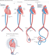
Fig. 1
Fig. 1. Left ventricular circulatory support devices.
a, The intra-aortic balloon pump uses counterpulsation to provide circulatory support and is commonly inserted via the femoral or axillary artery. b, Microaxial flow devices traverse the aortic valve via the femoral or axillary artery and continuously remove blood from the left ventricular (LV) cavity to achieve unloading. c, Transseptal percutaneous assist devices are powered by a centrifugal pump and are inserted via the femoral vein, across the intra-atrial septum and into the left atrium, with an outflow cannula in the femoral artery. Devices using centrifugal pumps require a separate control and monitoring module. d, Venoarterial extracorporeal membrane oxygenation uses a centrifugal pump to pull venous blood from the right atrium, through an oxygenator and into the arterial circulation via the outflow cannula in the aorta. Common cannulation sites include the femoral or axillary vessels or via central cannulation by thoracotomy or sternotomy. Extracorporeal membrane oxygenation can be used for left, right or biventricular support. e, Surgical extracorporeal centrifugal devices provide LV support via sternotomy or minimally invasive thoracotomy. Inflow cannulation is from the left ventricle or left atrium, and outflow cannulation is into the aorta.
Fig. 2
Fig. 2. Right ventricular circulatory support devices.
a, Microaxial flow devices for right ventricular (RV) support are inserted percutaneously through the femoral vein and into the pulmonary artery. b, Dual-lumen percutaneous assist devices draw blood from the right atrium or right ventricle to an external continuous centrifugal pump and out to the pulmonary artery. c, Percutaneous cannulas draw blood from the right atrium to a continuous centrifugal pump and into the pulmonary artery via the femoral, internal jugular or subclavian vein. d, Surgical extracorporeal centrifugal devices provide RV support via sternotomy or minimally invasive thoracotomy. Inflow cannulation is from the right atrium or right ventricle and outflow cannulation is into the pulmonary artery.
Fig. 3
Fig. 3. Effects of MCS devices on pressure–volume loops and blood pressure.
All temporary mechanical circulatory support (MCS) devices improve cardiac output (left) and blood pressure (right) to varying degrees. In this figure, the primary haemodynamic effects of temporary MCS devices are compared to illustrate the fundamental differences in how the devices interact with the heart and vasculature. Red lines indicate tracings under baseline cardiogenic shock conditions; blue lines show tracings after the introduction of the specified MCS device. a, Intra-aortic balloon pump (IABP). The tracings show the effects of counterpulsation, with a small reduction in left ventricular (LV) preload and a small increase in stroke volume. b, Microaxial flow pumps (connecting the left ventricle to the aorta). The tracings show substantial unloading (decreased LV end-diastolic pressure and pulmonary capillary wedge pressure), triangulation of the pressure–volume loop (indicating loss of isovolumic periods), decreased pressure–volume area (correlating with decreased myocardial oxygen consumption) and LV–aortic pressure uncoupling with a closed aortic valve. c, TandemHeart (connecting the left atrium to the femoral artery). The tracings show LV unloading (decreased LV end-diastolic pressure and pulmonary capillary wedge pressure), decreased LV stroke volume, increased aortic pressure (due to overall increased systemic blood flow), decreased pressure–volume area and preserved aortic valve opening (crucial for preventing LV and aortic root stasis and thrombosis). d, Peripheral extracorporeal membrane oxygenation (ECMO; connecting the right atrium to the femoral artery). The tracings show increasing LV preload (increased LV end-diastolic pressure and pulmonary capillary wedge pressure), decreased LV stroke volume, increased aortic pressure (due to overall increased systemic blood flow), increased pressure–volume area (correlating with increased myocardial oxygen consumption) and preserved aortic valve opening (crucial for preventing LV and aortic root stasis and thrombosis). e, Right-sided microaxial and extracorporeal centrifugal devices (connecting the right atrium to the pulmonary artery). The tracings show right ventricular (RV) unloading (decreased RV end-diastolic pressure and central venous pressure), decreased RV stroke volume, increased pulmonary artery pressure (due to overall increased pulmonary blood flow), decreased RV pressure–volume area and preserved pulmonary valve opening (crucial for preventing RV and pulmonary artery root stasis and thrombosis). All tracings were obtained using the Harvi cardiovascular simulation and are used with permission from PVLoops.

References

    1. Parissis H, et al. IABP: history-evolution-pathophysiology-indications: what we need to know. J. Cardiothorac. Surg. 2016;11:122. doi: 10.1186/s13019-016-0513-0. - DOI - PMC - PubMed
    1. Krishna M, Zacharowski K. Principles of intra-aortic balloon pump counterpulsation. Contin. Educ. Anaesth. Crit. Care Pain. 2009;9:24–28. doi: 10.1093/bjaceaccp/mkn051. - DOI
    1. Getinge. Intra-aortic balloon counterpulsation. Getingehttps://www.getinge.com/me/products/hospital/counterpulsation/ (2022).
    1. Burkhoff D, Sayer G, Doshi D, Uriel N. Hemodynamics of mechanical circulatory support. J. Am. Coll. Cardiol. 2015;66:2663–2674. doi: 10.1016/j.jacc.2015.10.017. - DOI - PubMed
    1. Schreuder JJ, et al. Beat-to-beat effects of intraaortic balloon pump timing on left ventricular performance in patients with low ejection fraction. Ann. Thorac. Surg. 2005;79:872–880. doi: 10.1016/j.athoracsur.2004.07.073. - DOI - PubMed