. 2022 Nov 6;11(11):1566.
doi: 10.3390/antibiotics11111566.

Phage Cocktail in Combination with Kasugamycin as a Potential Treatment for Fire Blight Caused by Erwinia amylovora

Affiliations

Phage Cocktail in Combination with Kasugamycin as a Potential Treatment for Fire Blight Caused by Erwinia amylovora

Sang-Guen Kim et al. Antibiotics (Basel). .

Abstract

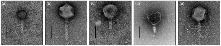
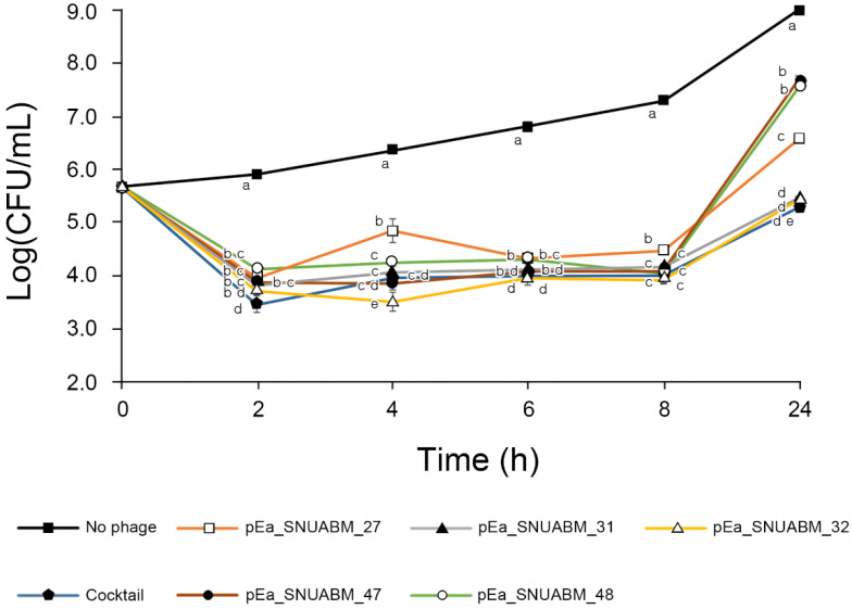
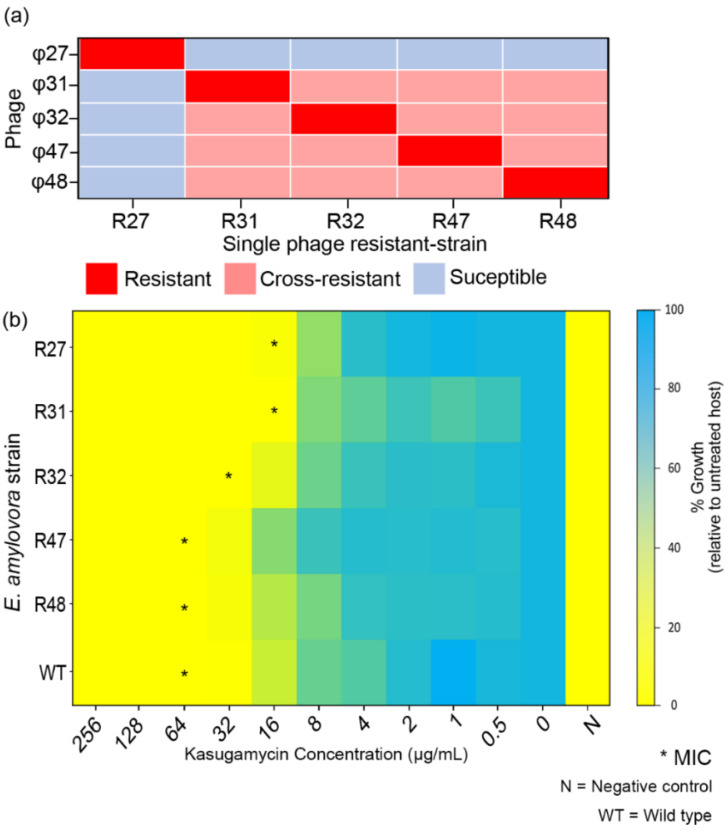
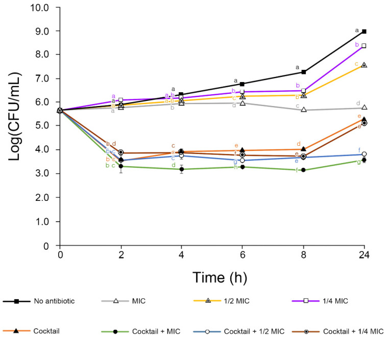
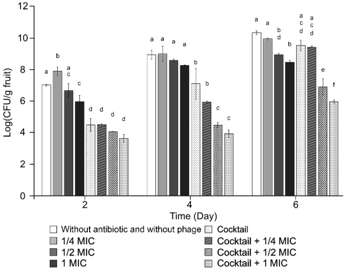
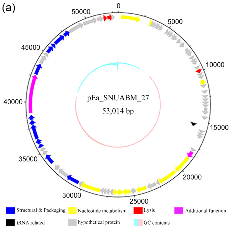
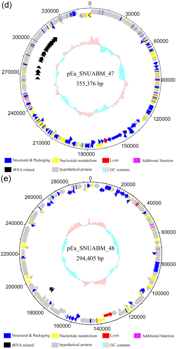
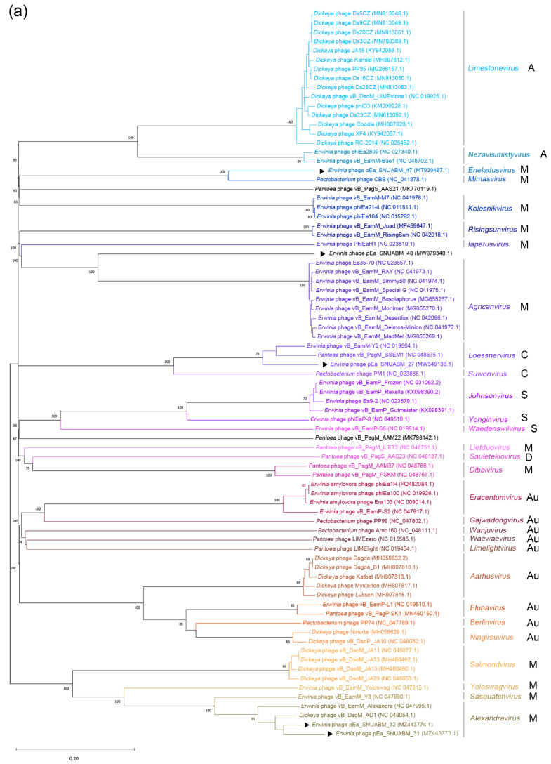
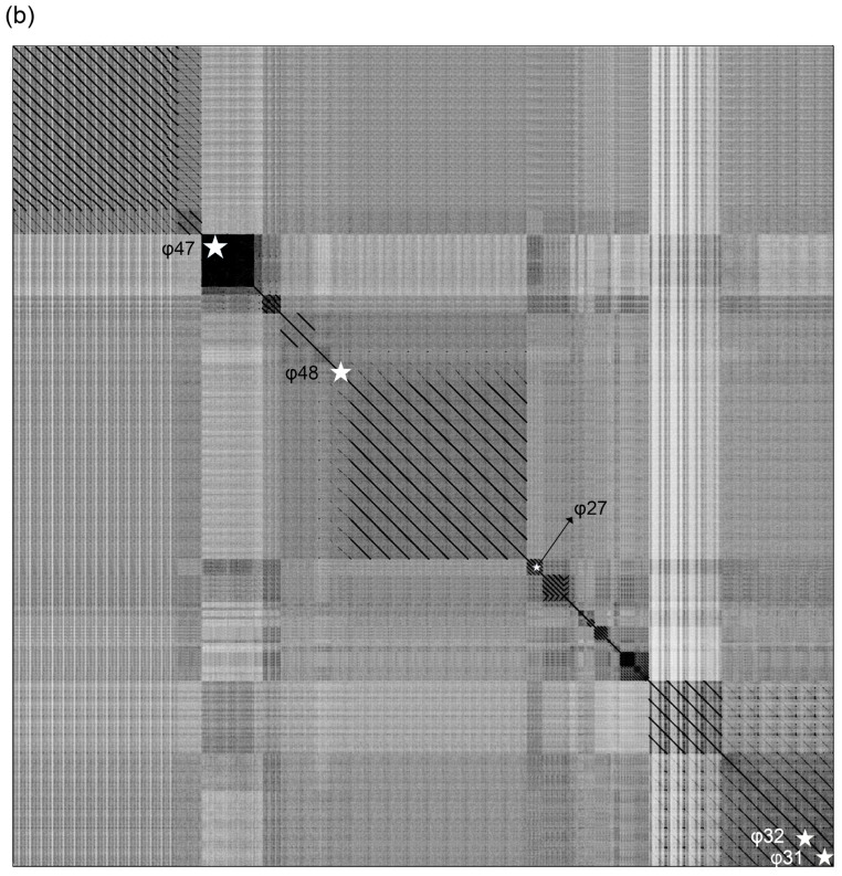
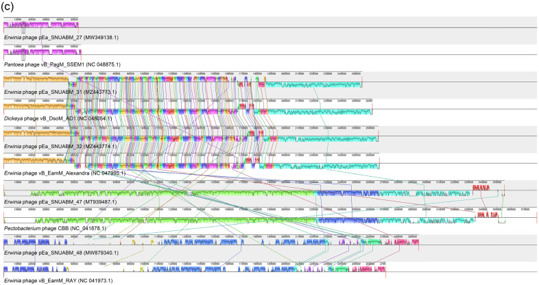
Recently, there has been an increasing number of blight disease reports associated with Erwinia amylovora and Erwinia pyrifoliae in South Korea. Current management protocols that have been conducted with antibiotics have faced resistance problems and the outbreak has not decreased. Because of this concern, the present study aimed to provide an alternative method to control the invasive fire blight outbreak in the nation using bacteriophages (phages) in combination with an antibiotic agent (kasugamycin). Among 54 phage isolates, we selected five phages, pEa_SNUABM_27, 31, 32, 47, and 48, based on their bacteriolytic efficacy. Although only phage pEa_SNUABM_27 showed host specificity for E. amylovora, all five phages presented complementary lytic potential that improved the host infectivity coverage of each phage All the phages in the cocktail solution could lyse phage-resistant strains. These strains had a decreased tolerance to the antibiotic kasugamycin, and a synergistic effect of phages and antibiotics was demonstrated both in vitro and on immature wound-infected apples. It is noteworthy that the antibacterial effect of the phage cocktail or phage cocktail-sub-minimal inhibitory concentration (MIC) of kasugamycin was significantly higher than the kasugamycin at the MIC. The selected phages were experimentally stable under environmental factors such as thermal or pH stress. Genomic analysis revealed these are novel Erwinia-infecting phages, and did not encode antibiotic-, virulence-, or lysogenic phage-related genes. In conclusion, we suggest the potential of the phage cocktail and kasugamycin combination as an effective strategy that would minimize the use of antibiotics, which are being excessively used in order to control fire blight pathogens.

Keywords: Erwinia amylovora; bacteriophage; fire blight; phage therapy; phage–antibiotic synergy.


Conflict of interest statement

The authors declare no conflict of interest.

Figures

Figure 1
Figure 1
Transmission electron micrographs of Erwinia bacteriophages (a) φ27, (b) φ31, (c) φ32, (d) φ47, and (e) φ48. Scale bar is 100 nm. The contractile tails of φ32, φ47, and φ48 were observed in the contracted state (ce).
Figure 2
Figure 2
In vitro bactericidal effect of Erwinia phages φ27, φ31, φ32, φ47, φ48, and their cocktail. The viable bacterial cells were counted over 24 h. The E. amylovora strain TS3128, a reference strain for research in Korea, was used. The bars of each point indicate the standard deviation. Statistical significance was calculated using the one-way analysis of variance test with Tukey post-hoc, and the significance threshold was set at p < 0.05. Means at the same sampling time point with different letters (a–e) are significantly different.
Figure 3
Figure 3
Biological characteristics of phage-resistant Erwinia amylovora TS3128. (a) Phage resistance profiles of the single phage-resistant strains. (b) Minimum inhibitory concentration (MIC) of kasugamycin with the phage-resistant strains is indicated (*). WT and N indicate wild type and negative control (no bacterial ingredient), respectively.
Figure 4
Figure 4
In vitro phage cocktail–antibiotic synergy assay with Erwinia amylovora TS3128. The viable bacterial cells were counted over 24 h. The bars of each point indicate the standard deviation. Statistical significance was calculated using the one-way analysis of variance test with Tukey post-hoc, and the significance threshold was set at p < 0.05. Means at the same sampling time point with different letters (a–g) are significantly different.
Figure 5
Figure 5
Apple fruit administration of the five-phage cocktail in combination with 0, 1/4, 1/2, or 1 MIC kasugamycin, under controlled conditions. The infective concentration of Erwinia amylovora TS3128 was 2 × 10⁵ Colony Forming Unit [CFU]/mL. Viable bacterial cell counts were observed over time. The bars of each point indicate the standard deviation. Statistical significance was calculated using the one-way analysis of variance test with Tukey post-hoc, and the significance threshold was set at p < 0.05. Means at the same sampling time point with different letters (a–f) are significantly different.
Figure 6
Figure 6
Genome map of Erwinia phages (a) φ27, (b) φ31, (c) φ32, (d) φ47, and (e) φ48. The open reading frames were functionally assorted into six groups of proteins related to: structure and packaging (blue), nucleotide metabolism (yellow), lysis (red), and additional functions (purple), as well as tRNA proteins or tRNA-related proteins (black), and hypothetical proteins (gray). Scale is base pair (bp).
Figure 6
Figure 6
Genome map of Erwinia phages (a) φ27, (b) φ31, (c) φ32, (d) φ47, and (e) φ48. The open reading frames were functionally assorted into six groups of proteins related to: structure and packaging (blue), nucleotide metabolism (yellow), lysis (red), and additional functions (purple), as well as tRNA proteins or tRNA-related proteins (black), and hypothetical proteins (gray). Scale is base pair (bp).
Figure 7
Figure 7
Phylogenetic whole genome analysis of 79 phages infecting Erwinia spp., Dickeya spp., Pantoea spp., and Pectobacterium spp. (a) The phylogenetic tree was constructed using Virus Classification and Tree Building Online Resource (VICTOR). Black arrows (▶) indicate the five Erwinia phages in this study. Black letters next to genus indicate family of the phages (A: Ackermannviridae, M: Myoviridae, C: Chaseviridae, S: Schitoviridae, D: Drexlerviridae, Au: Autographiviridae). The genus of φ27 was identified as Loessnervirus, that of φ31 and φ32 as Alexandravirus, that of φ47 as Eneladusvirus, while that of φ48 was unclassified. (b) Dot plot analysis of the 79 phages with parallel order of phylogeny. (c) Comparative whole genome analysis using progressive Mauve.
Figure 7
Figure 7
Phylogenetic whole genome analysis of 79 phages infecting Erwinia spp., Dickeya spp., Pantoea spp., and Pectobacterium spp. (a) The phylogenetic tree was constructed using Virus Classification and Tree Building Online Resource (VICTOR). Black arrows (▶) indicate the five Erwinia phages in this study. Black letters next to genus indicate family of the phages (A: Ackermannviridae, M: Myoviridae, C: Chaseviridae, S: Schitoviridae, D: Drexlerviridae, Au: Autographiviridae). The genus of φ27 was identified as Loessnervirus, that of φ31 and φ32 as Alexandravirus, that of φ47 as Eneladusvirus, while that of φ48 was unclassified. (b) Dot plot analysis of the 79 phages with parallel order of phylogeny. (c) Comparative whole genome analysis using progressive Mauve.

References

    1. Llop P., Bonaterra A., Peñalver J., López M.M. Development of a highly sensitive nested-PCR procedure using a single closed tube for detection of Erwinia amylovora in asymptomatic plant material. Appl. Environ. Microbiol. 2000;66:2071–2078. doi: 10.1128/AEM.66.5.2071-2078.2000. - DOI - PMC - PubMed
    1. Kharadi R.R., Castiblanco L.F., Waters C.M., Sundin G.W. Phosphodiesterase genes regulate amylovoran production, biofilm formation, and virulence in Erwinia amylovora. Appl. Environ. Microbiol. 2019;85:e02233-18. doi: 10.1128/AEM.02233-18. - DOI - PMC - PubMed
    1. Adaskaveg J.E., Förster H., Wade M.L. Effectiveness of kasugamycin against Erwinia amylovora and its potential use for managing fire blight of pear. Plant Dis. 2011;95:448–454. doi: 10.1094/PDIS-09-10-0679. - DOI - PubMed
    1. Jurgens A.G., Babadoost M. Sensitivity of Erwinia amylovora in Illinois apple orchards to streptomycin, oxytetracyline, kasugamycin, and copper. Plant Dis. 2013;97:1484–1490. doi: 10.1094/PDIS-02-13-0209-RE. - DOI - PubMed
    1. Aćimović S.G., Zeng Q., McGhee G.C., Sundin G.W., Wise J.C. Control of fire blight (Erwinia amylovora) on apple trees with trunk-injected plant resistance inducers and antibiotics and assessment of induction of pathogenesis-related protein genes. Front. Plant Sci. 2015;6:16. - PMC - PubMed

LinkOut - more resources