. 2022 Nov 1;12(11):1484.
doi: 10.3390/brainsci12111484.

Multiple Machine Learning Approaches for Morphometric Parameters in Prediction of Hydrocephalus

Affiliations

Multiple Machine Learning Approaches for Morphometric Parameters in Prediction of Hydrocephalus

Hao Xu et al. Brain Sci. .

Abstract

Background: The diagnosis of hydrocephalus is mainly based on imaging findings. However, the significance of many imaging indicators may change, especially in some degenerative diseases, and even lead to misdiagnosis.

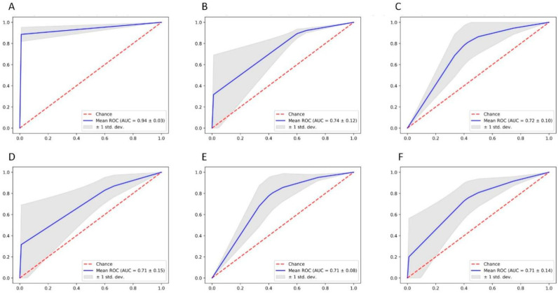
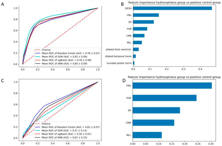
Methods: This study explored the effectiveness of commonly used morphological parameters and typical radiographic findings in hydrocephalus diagnosis. The patients' imaging data were divided into three groups, including the hydrocephalus group, the symptomatic group, and the normal control group. The diagnostic validity and weight of various parameters were compared between groups by multiple machine learning methods.

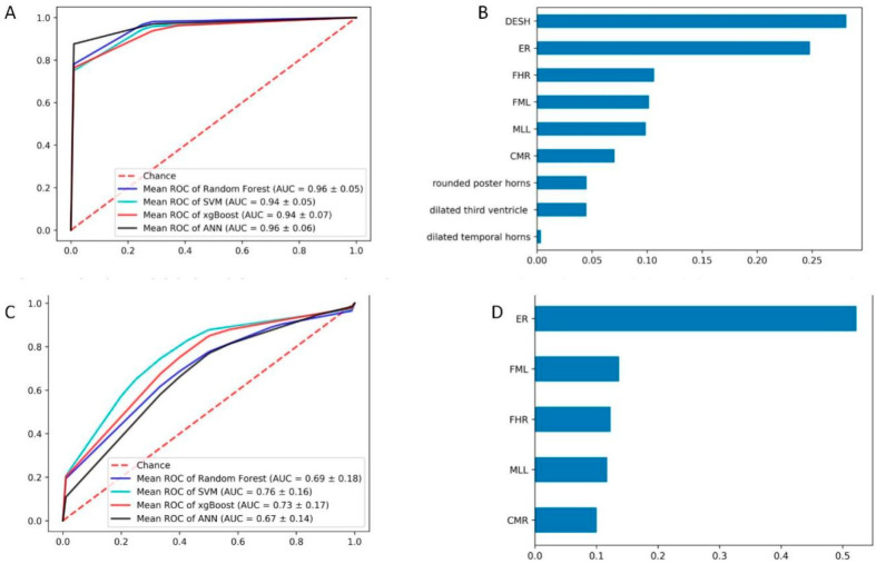
Results: Our results demonstrated that Evans' ratio is the most valuable diagnostic indicator compared to the hydrocephalus group and the normal control group. But frontal horns' ratio is more useful in diagnosing patients with symptoms. Meanwhile, the sign of disproportionately enlarged subarachnoid space and third ventricle enlargement could be effective diagnostic indicators in all situations.

Conclusion: Both morphometric parameters and radiological features were essential in diagnosing hydrocephalus, but the weights are different in different situations. The machine learning approaches can be applied to optimize the diagnosis of other diseases and consistently update the clinical diagnostic criteria.

Keywords: hydrocephalus; imaging diagnosis; machine learning.


Conflict of interest statement

We confirm that the manuscript has been read and approved by all named authors and that there are no other persons who satisfied the criteria for authorship but are not listed. We further confirm that the order of authors listed in the manuscript has been approved by all of us. The authors declare no conflict of interest.

Figures

Figure 1
Figure 1
Radiological features and morphometric parameters in CT image. (A). The layer of enlargement ventricle; (B). The layer of third ventricle; (C). The layer of maximum width of the internal diameter (D). The layer of maximum transverse diameter of the skull and the narrowest width between the lateral walls (not necessarily on same level). a. Dilated temporal horns; b. Rounded posterior horns; c. Dilated third ventricle; d. Frontal Horns’ Length; e, Inner diameter of the skull in the same line as FHL; f. Maximum width of the internal diameter of the skull; g. The narrowest width between the lateral walls; h. Maximum transverse diameter of the skull.
Figure 2
Figure 2
Training and evaluation procedures of the machine learning model.
Figure 3
Figure 3
The comparison of hydrocephalus group and symptomatic group in terms of ROC and weight. (A) ROC curves of the four derived models with radiological features; RF, random forest, XG, XGBoost; ANN, artificial neural network; SVM, support vector machine for the patients with hydrocephalus and normal control; (B) The weight of all features; (C) ROC curves of the four derived models with morphometric parameters; (D) The weight of all features morphometric parameters.
Figure 4
Figure 4
The ROC of confusion matrix diagnosis index in the comparison of hydrocephalus group and symptomatic group. (A) dilated temporal horns, dilated third ventricle, rounded posterior horns, and DESH; (B) ER + FHR; (C) FHR + CMR; (D) ER + FHR + CMR; (E) FHR + CMR; (F) ER.
Figure 5
Figure 5
The comparison of hydrocephalus group and symptomatic group in terms of ROC and weight. (A) ROC curves of the four derived models with all features: RF, random forest, XG, XGBoost; ANN, artificial neural network; SVM, support vector machine for the patients with hydrocephalus and normal control; (B) The weight of all features; (C) ROC curves of the four derived models with morphometric parameters; (D) The weight of all features morphometric parameters.

Similar articles

Cited by

References

    1. Kahle K.T., Kulkarni A.V., Limbrick D.D., Jr., Warf B.C. Hydrocephalus in children. Lancet. 2016;387:788–799. doi: 10.1016/S0140-6736(15)60694-8. - DOI - PubMed
    1. Symss N.P., Oi S. Theories of cerebrospinal fluid dynamics and hydrocephalus: Historical trend. J. Neurosurg. Pediatr. 2013;11:170–177. doi: 10.3171/2012.3.PEDS0934. - DOI - PubMed
    1. Pisapia J.M., Rozycki M., Akbari H., Bakas S., Thawani J.P., Moldenhauer J.S., Storm P.B., Zarnow D.M., Davatzikos C., Heuer G.G. Correlations of atrial diameter and frontooccipital horn ratio with ventricle size in fetal ventriculomegaly. J. Neurosurg. Pediatr. 2017;19:300–306. doi: 10.3171/2016.9.PEDS16210. - DOI - PubMed
    1. Zhuravlova I., Kornieieva M.J. Anatomic Variability of the Morphometric Parameters of the Third Ventricle of the Brain and Its Relations to the Shape of the Skull. Neurol. Surg. B Skull Base. 2021;82((Suppl. S3)):e315–e320. doi: 10.1055/s-0040-1701527. - DOI - PMC - PubMed
    1. Friedman D.I., Jacobson D.M. Diagnostic criteria for idiopathic intracranial hypertension. Neurology. 2002;59:1492–1495. doi: 10.1212/01.WNL.0000029570.69134.1B. - DOI - PubMed

LinkOut - more resources