. 2022 Oct 28;11(11):2133.
doi: 10.3390/antiox11112133.

Quercetin Abates Aluminum Trioxide Nanoparticles and Lead Acetate Induced Altered Sperm Quality, Testicular Oxidative Damage, and Sexual Hormones Disruption in Male Rats

Affiliations

Quercetin Abates Aluminum Trioxide Nanoparticles and Lead Acetate Induced Altered Sperm Quality, Testicular Oxidative Damage, and Sexual Hormones Disruption in Male Rats

Amany Behairy et al. Antioxidants (Basel). .

Abstract

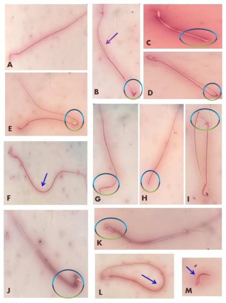
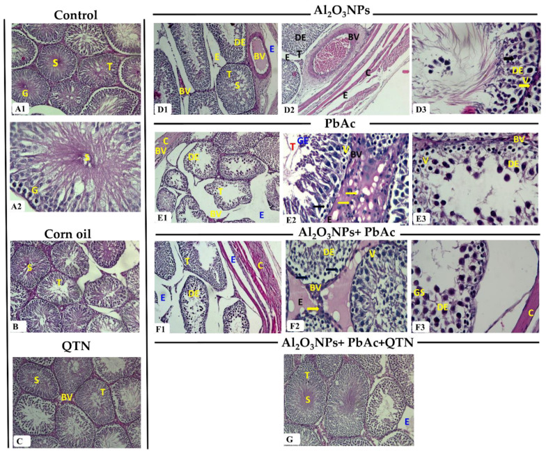
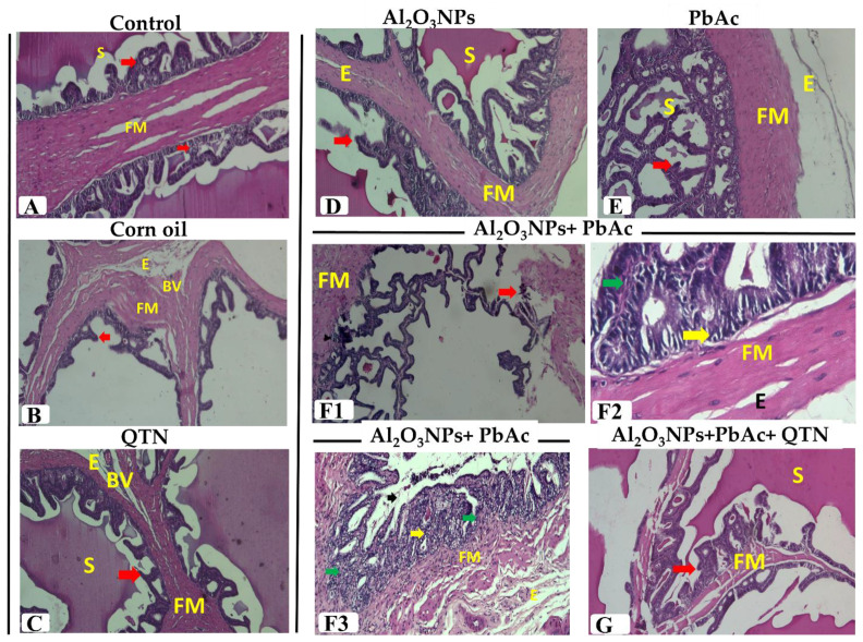
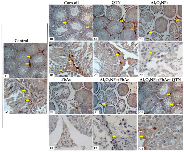
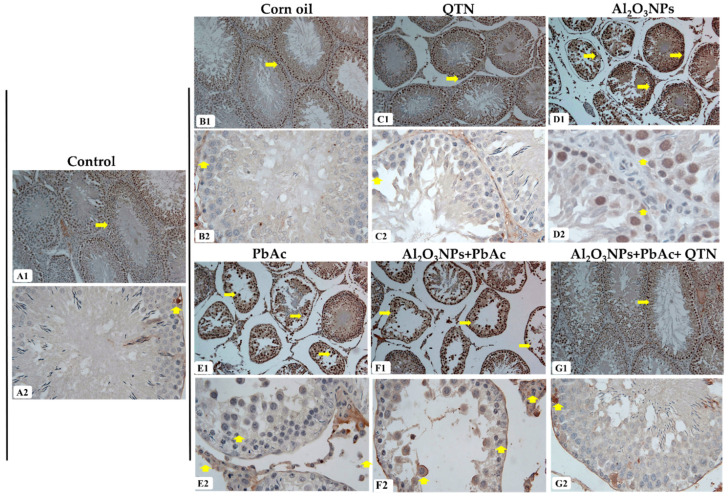
This study examined the effects of exposure to lead acetate (PbAc) and/or aluminum trioxide nanoparticles (Al2O3NPs) on testicular function. Additionally, the probable reproprotective effects of quercetin (QTN) against Al2O3NPs and PbAc co-exposure in male Sprague Dawely rats were assessed. Al2O3NPs (100 mg/kg b.wt.), PbAc (50 mg/kg b.wt.), and QTN (20 mg/kg b.wt.) were orally administered for 60 days. Then, spermiogram, histopathological examinations of the testis and accessory glands, and immunohistochemical detection of androgen receptors (AR) and tumor necrotic factor alpha (TNF-α) were achieved. Moreover, serum levels of male sex hormones and testicular levels of antioxidant indices were estimated. The results showed that Al2O3NPs and/or PbAc caused significant sperm abnormalities, testicular oxidative stress, and histopathological changes. Furthermore, serum testosterone, LH, and FSH levels significantly decreased, while estradiol levels significantly increased. The Al2O3NPs and/or PbAc co-exposed group had more obvious disturbances. Furthermore, QTN co-administration significantly reversed the Al2O3NPs and PbAc-induced testicular histopathological alterations, reduced antioxidant defenses, and altered AR and TNF-α immune expression in testicular tissues. Conclusively, Al2O3NPs and/or PbAc evoked testicular dysfunction by inducing oxidative injury and inflammation. However, QTN oral dosing effectively mitigated the negative effects of Al2O3NPs and PbAc by suppressing oxidative stress and inflammation and improving the antioxidant defense system.

Keywords: aluminum trioxide nanoparticles; androgen receptors; inflammation; lead acetate; oxidative stress; quercetin; rats; tumor necrotic factor alpha.


Conflict of interest statement

The authors declare no conflict of interest.

Figures

Figure 1
Figure 1
Abnormal spermatozoa induced in rats exposed to Al2O3NPs and/or PbAc. (A) Normal sperm, (B) amorphous head with double tail, (C) short tail, (D,E) bent tail, (F) curved tail, (G) bent head, (H) detached head, (I) broken tail, (J) broken head, (K) flattened head, (L) looped tail, (M) detached tail.
Figure 2
Figure 2
(A1) Testis of control group showing normal structure. It consisted of seminiferous tubules (T) lined by stratified germinal epithelium (G) and lumen filled with spermatozoa (S) (H&E, ×100). (A2) Testis of the control group showing stratified germinal epithelium (G) lined seminiferous tubules (T) and spermatozoa (S) in the tubular lumen (H&E, ×400). (B) Testis of corn oil-treated group showing normal seminiferous tubules (T) lined by several layers of germinal epithelium (G) and spermatozoa (S) in the lumen (H&E, ×100). (C) Testis of QTN-treated group showing several rows of germinal epithelium (G) lined seminiferous tubules (T) and spermatozoa (S) in the lumen (H&E, ×100). (D1) Testis of Al2O3NPs-exposed group showing atrophied seminiferous tubules (T) with few spermatozoa (S) in the lumen, disorganized epithelium (DE), severe congestion of the interstitial and sub-capsular blood vessels (BV) and edema (E) (H&E, ×100). (D2) Testis of Al2O3NPs-exposed group showing disorganized epithelium (DE) in the seminiferous tubules (T), interstitial and capsular edema (E), severe congestion of the thickened wall sub-capsular blood vessel (BV) as well as a thick capsule (C) (H&E, ×100). (D3) Testis of Al2O3NPs-exposed group showing epithelial disorganization (DE), sloughing of germinal epithelium, necrotic epithelium, and darkly stained pyknotic nuclei (Black arrow), the vacuolated epithelium (V), thickened basement membrane (Yellow arrow) and few spermatozoa (S) in lumen (H&E, ×400). (E1) Testis of PbAc-exposed group showing atrophied seminiferous tubules (T) with a corrugated outline, sloughed germinal epithelium, the disorganized epithelium (DE), severe congestion of interstitial and sub-capsular blood vessels (BV), edema (E), and thickened capsule (C) (H&E, ×100). (E2) Testis of PbAc-exposed group showing sloughed germinal epithelium (GE) within seminiferous tubules (T), necrotic with darkly stained pyknotic nuclei (Arrow) and vacuolated (V) germinal epithelium, congestion of interstitial blood vessels (BV), edema (E), and interstitial inflammatory cell infiltrations (yellow arrow) (H&E, ×400). (E3) Testis of PbAc-exposed group showing disorganized (DE), sloughed, necrotic and vacuolated (V) germinal epithelium, and congested blood vessel (BV) (H&E, ×400). (F1) Testis of Al2O3NPs and PbAc co-exposed group showing shrunken tubules with different shapes and irregular outlines, germinal disorganization (DE), loss of the germinal epithelium, edema (E), and thick capsule (C) (H&E, ×100). (F2) Testis of Al2O3NPs and PbAc co-exposed group showing germinal disorganization (DE), necrotic with darkly stained pyknotic nuclei (Arrow), cellular vacuolation (V), congestion of interstitial blood vessels (BV), edema (E), and interstitial inflammatory cell infiltrations (yellow arrow) (H&E, ×400). (F3) Testis of Al2O3NPs and PbAc co-exposed group showing epithelial disorganization (DE), cellular vacuolation (V), giant spermatids (GS), subcapsular edema (E), and thick capsule (C) (H&E, ×400). (G) Testis of Al2O3NPs and PbAc co-exposure and QTN-treated group showing stratified germinal epithelium-lined seminiferous tubules (T), spermatozoa (S) in the lumen, and mild focal edema (E) (H&E, ×100).
Figure 3
Figure 3
(A) Prostate gland of the control group showing normal structure as different sizes and shapes acini (A) with prostatic secretion (S) in the lumen (H&E, ×100). (B) Prostate gland of corn oil-treated group showing normal structure with normal acini (A) and secretion (S) in the lumen (H&E, ×40). (C) Prostate gland of QTN-treated group showing normal acini (A) and secretion (S) in the lumen (H&E, ×100). (D) Prostate gland of Al2O3NPs-exposed group showing hyperplasia of acini (A) with papillary folds (Red arrow) and scanty secretion (S) in the lumen (H&E, ×100). (E1) Prostate gland of PbAc-exposed group showing prostatic acinar hyperplasia with papillary folds (Red arrow), destructed acini and decreasing acinar size (A), interstitial inflammatory cells and congested blood vessels (BV), thick fibro-muscular stroma (FM) as well as little secretion (S) (H&E, ×100). (E2) Prostate gland of PbAc-exposed group showing prostatic acinar hyperplasia with papillary folds (Red arrow), destructed acini and decreasing acinar size (A), interstitial inflammatory cells (Yellow arrow) and thick fibro-muscular stroma (FM) as well as little secretion (S) (H&E, ×100). (F1) Prostate gland of Al2O3NPs and PbAc co-exposed group showing marked prostatic acinar hyperplasia with papillary folds (Red arrow), decreasing acinar size, destructed acini (A), thick fibro-muscular stroma (FM), and little secretion (S) (H&E, ×100). (F2) Prostate gland of Al2O3NPs and PbAc co-exposed group showing acini lined with vacuolated epithelium, acinar hyperplasia with papillary folds (Red arrow), destructed acini, thick fibro-muscular stroma (FM), few secretions (S), inflammatory cells around the acini and congested blood vessels (BV) (H&E, ×400). (F3) Prostate gland of Al2O3NPs and PbAc co-exposed group showing prostatic acinar with secretion (S) and inflammatory cells (Yellow arrow) around the acini (H&E, ×400). (G) Prostate gland of Al2O3NPs and PbAc co-exposure and QTN-treated group showing normal structure with normal acini (A), secretion (S) in the lumen, and very mild edema (E) (H&E, ×100).
Figure 4
Figure 4
(A) Seminal vesicle of control group showing normal branched mucosal folds (Red arrow) surrounded by fibro-muscular layer (FM) and secretion (S) in the lumen (H&E, ×100). (B) Seminal vesicle of corn oil-treated group showing mucosal folds (Red arrow) as well as edema (E) and congestion in the blood vessel (BV) in fibro-muscular layer (FM) (H&E, ×100). (C) Seminal vesicle of QTN-treated group showing normal structure as mucosal folds (Red arrow) and secretion (S) in the lumen as well as edema (E) and congested blood vessel (BV) in fibro-muscular layer (FM) (H&E, ×100). (D) Seminal vesicle of Al2O3NPs-exposed group showing hyperplastic epithelial folds (Red arrow), little secretion (S) in the lumen and thickened fibro-muscular layer (FM) with edema (E) (H&E, ×100). (E) Seminal vesicle of PbAc-exposed group showing hyperplastic epithelial folds (Red arrow), few secretions (S), subcapsular edema (E), and thickened fibro-muscular layer (FM) (H&E, ×100). (F1) Seminal vesicle of Al2O3NPs and PbAc co-exposed group showing destructed folds (Red arrow) and thickened fibro-muscular layer (FM) (H&E, ×100). (F2) Seminal vesicle of Al2O3NPs and PbAc co-exposed group showing vacuolated epithelium (Yellow arrow), interstitial mononuclear inflammatory cells (Green arrow), and thickened fibro-muscular layer (FM) with edema (E) (H&E, ×400). (F3) Seminal vesicle of Al2O3NPs and PbAc co-exposed group showing destructed folds (arrow) vacuolated epithelium (Yellow arrow), mononuclear inflammatory cells (Green arrow), thickened fibro-muscular layer (FM), and edema (E) (H&E, ×100). (G) Seminal vesicle of Al2O3NPs and PbAc co-exposure and QTN-treated group showing normal mucosal folds (Red arrow) surrounded by fibro-muscular layer (FM) and secretions (S) in the lumen (H&E, ×100).
Figure 5
Figure 5
(A1,A2) Testis of the control group showing strong interstitial cell staining for androgen receptors and lack of immuno-reaction in seminiferous tubules. (B1,B2) Testis of corn oil-treated group showing normal immuno-expression in the interstitial cells. (C1,C2) Testis of QTN-treated group showing normal expression of immuno-staining of interstitial cells. (D1,D2) Testis of Al2O3NPs-exposed group showing moderate immuno-expression of the androgen receptor. (E1,E2) Testis of PbAc-exposed group showing weak androgen receptor expression. (F1,F2) Testis of Al2O3NPs and PbAc co-exposed group showing weak androgen receptor expression. (G1,G2) Testis of Al2O3NPs and PbAc co-exposed and QTN-treated group showing strong androgen receptor expression in the interstitial cells (Androgen receptor immunostain, ×100 for (A1G1) and ×400 (A2G2)). Yellow arrows denoted AR-positive immune stained cells.
Figure 6
Figure 6
(A1,A2) Testis of the control rat showing weak TNF-α immuno-stain in the cytoplasm of few spermatogenic cells. (B1,B2) Testis of corn oil-treated rat showing weak expression for TNF-α. (C1,C2) Testis of QTN-treated rat showing weak immuno-labeling of some spermatogenic cells. (D1,D2) Testis of Al2O3NPs-exposed group showing numerous positive spermatogenic cells and Leydig cells represented by brown color. (E1,E2) Testis of the PbAc-exposed group showing a strong positive reaction in the cytoplasm of spermatogenic cells and Leydig cells. (F1,F2) Testis of Al2O3NPs and PbAc co-exposed group showing strong positive TNF-α immuno-stain (G1,G2) Testis of Al2O3NPs and PbAc co-exposed and QTN-treated group showing weak TNF-α positive cells (TNF-α immuno-stained, ×100 for (A1G1) and ×400 (A2G2)). Yellow arrows denoted TNF-α-positive immune stained cells.

Similar articles

Cited by

References

    1. Abd-Elhakim Y.M., Hashem M.M., Abo-El-Sooud K., Hassan B.A., Elbohi K.M., Al-Sagheer A.A. Effects of Co-Exposure of Nanoparticles and Metals on Different Organisms: A Review. Toxics. 2021;9:284. doi: 10.3390/toxics9110284. - DOI - PMC - PubMed
    1. Dantas G.P.F., Ferraz F.S., Andrade L.M., Costa G.M.J. Male reproductive toxicity of inorganic nanoparticles in rodent models: A systematic review. Chem.-Biol. Interact. 2022;363:110023. doi: 10.1016/j.cbi.2022.110023. - DOI - PubMed
    1. Ni D.Q., Ma D.D., Hao S.L., Yang W.X., Kovacs T., Tan F.Q. Titanium dioxide nanoparticles perturb the blood-testis barrier via disruption of actin-based cell adhesive function. Aging. 2021;13:25440–25452. doi: 10.18632/aging.203763. - DOI - PMC - PubMed
    1. Asare N., Instanes C., Sandberg W.J., Refsnes M., Schwarze P., Kruszewski M., Brunborg G. Cytotoxic and genotoxic effects of silver nanoparticles in testicular cells. Toxicology. 2012;291:65–72. doi: 10.1016/j.tox.2011.10.022. - DOI - PubMed
    1. Hamdi H. Testicular dysfunction induced by aluminum oxide nanoparticle administration in albino rats and the possible protective role of the pumpkin seed oil. J. Basic Appl. Zool. 2020;81:42. doi: 10.1186/s41936-020-00178-8. - DOI