Review
. 2022 Oct 29;11(11):2143.
doi: 10.3390/antiox11112143.

Roles of Ferredoxin-NADP+ Oxidoreductase and Flavodoxin in NAD(P)H-Dependent Electron Transfer Systems

Affiliations
Review

Roles of Ferredoxin-NADP+ Oxidoreductase and Flavodoxin in NAD(P)H-Dependent Electron Transfer Systems

Takashi Iyanagi. Antioxidants (Basel). .

Abstract

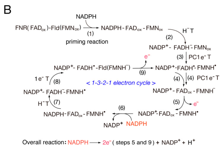
Distinct isoforms of FAD-containing ferredoxin-NADP+ oxidoreductase (FNR) and ferredoxin (Fd) are involved in photosynthetic and non-photosynthetic electron transfer systems. The FNR (FAD)-Fd [2Fe-2S] redox pair complex switches between one- and two-electron transfer reactions in steps involving FAD semiquinone intermediates. In cyanobacteria and some algae, one-electron carrier Fd serves as a substitute for low-potential FMN-containing flavodoxin (Fld) during growth under low-iron conditions. This complex evolves into the covalent FNR (FAD)-Fld (FMN) pair, which participates in a wide variety of NAD(P)H-dependent metabolic pathways as an electron donor, including bacterial sulfite reductase, cytochrome P450 BM3, plant or mammalian cytochrome P450 reductase and nitric oxide synthase isoforms. These electron transfer systems share the conserved Ser-Glu/Asp pair in the active site of the FAD module. In addition to physiological electron acceptors, the NAD(P)H-dependent diflavin reductase family catalyzes a one-electron reduction of artificial electron acceptors such as quinone-containing anticancer drugs. Conversely, NAD(P)H: quinone oxidoreductase (NQO1), which shares a Fld-like active site, functions as a typical two-electron transfer antioxidant enzyme, and the NQO1 and UDP-glucuronosyltransfease/sulfotransferase pairs function as an antioxidant detoxification system. In this review, the roles of the plant FNR-Fd and FNR-Fld complex pairs were compared to those of the diflavin reductase (FAD-FMN) family. In the final section, evolutionary aspects of NAD(P)H-dependent multi-domain electron transfer systems are discussed.

Keywords: catalytic cycle; diflavin reductase family; electron transfer; evolutionary aspects; ferredoxin (Fd); ferredoxin-NADP+ oxidoreductase (FNR); flavodoxin (Fld); redox potentials.


Conflict of interest statement

The author has no conflict of interest to declare.

Figures

Figure 1
Figure 1
Proposed catalytic cycles of the LFNR (A) and RFNR (B) systems. (A) Photosystem I → LFd I (2S-2Fe) → LFNR (FAD) → NADP+ [45] and (B) NADPH→RFNR (FAD) → RFd III→metabolic pathways. HT, hydride transfer; 1eT, one-electron transfer; PC1eT, proton-coupled one-electron transfer.
Scheme 1
Scheme 1
Switching between one-electron and two-electron transfer reactions in the FNR-Fd I/Fld and FNR-Fd III/Fld systems. HT, hydride transfer.
Figure 2
Figure 2
Proposed catalytic cycle of the FNR-Fld systems. (A) Photosystem I → Fld (FMN) → FNR (FAD) → NADP+ and (B) NADPH → FNR (FAD) → Fld (FMN) → metabolic redox pathways. In (B), the overall reaction is based on the catalytic cycle.
Figure 2
Figure 2
Proposed catalytic cycle of the FNR-Fld systems. (A) Photosystem I → Fld (FMN) → FNR (FAD) → NADP+ and (B) NADPH → FNR (FAD) → Fld (FMN) → metabolic redox pathways. In (B), the overall reaction is based on the catalytic cycle.
Figure 3
Figure 3
Proposed catalytic cycle of the eukaryotic-membrane-bound cyt P450 reductase-cyt P450 system. In this figure, the overall reaction is based on the catalytic cycle. Note that cyt P450R indicates cyt P450 reductase. HT, hydride transfer; 1eT, one-electron transfer; PC1e T, proton-coupled one-electron transfer.
Scheme 2
Scheme 2
The roles of FNR (FAD) and one-electron carriers in the NAD(P)H-dependent electron transfer systems.
Figure 4
Figure 4
Proposed catalytic cycle of NADH-cyt b5 reductase. Note that cytb5R (FADox) indicates oxidized cyt b5 reductase.
Figure 5
Figure 5
Proposed catalytic cycle for flavohemoglobin.
Figure 6
Figure 6
Membrane-bound NADPH-oxidase.
Figure 7
Figure 7
Proposed catalytic cycle of wild and mutant FNR Glu301Ala.
Figure 8
Figure 8
Proposed catalytic cycle of NQO1.
Figure 9
Figure 9
Evolutionary origin of the cyt P450 reductase: (left) plant FNR and bacterial Fld redox complex and (right) cyt P450 reductase. CD/H indicates the connecting domain (CD) and the hinge (H) [12,14].
Figure 10
Figure 10
Evolutionary model of the formation of the diflavin reductase family. All domains are arranged in order of the C-terminus (left) to the N-terminus (right), and the FNR (FAD) domain is positioned on the left. FNR-Fd indicates a fusion gene composed of FNR and Fld. sFNR-Fld indicates soluble ancestral FNR-Fld reductase. mP450R, membrane-bound cyt P450 reductase; FldR, flavodoxin reductase; SiFP, bacteria sulfite reductase flavoprotein component; P450BM3, cytochrome P450BM3 from Bacillus megaterium. For cyt 450 reductase and NOS isoforms, a dot indicates the conserved proximal cysteine residue, where the heme domains for the bacterial and mammalian NOS isoforms share a sequence motif, (R/K) C (I/V) G in the conserved proximal cysteine residues, which is found in cyt P450 gene family 1 [14]. The CD/H indicates the connecting (C) and hinge (H) domains (gray). TM indicates the membrane-binding domain. The electron transfer occurs from the electron donor NAD(P)H to the final electron acceptor heme iron: NAD(P)H → FAD → FMN/or [2Fe-2S] → Heme iron. HGT (red) indicates the transfer of the bacterial Fld gene to plant cells. HGT? (red) indicates the transfer of the plant FNR-Fld or sFNR-Fld gene to bacterial cells. Thus, the FAD-FMN module of bacterial SiR and P450BM3, mammalian cyt P450 reductase and NOS isoforms are derived from plant FNR-Fld or sFNR-Fld as a precursor gene [14,21].
Figure 11
Figure 11
Electron transfer cascades for plant sulfite reductase. Figure was adapted with permission from Refs. [132,133] with some modifications. Copyright year 2005, Hase et al. (2005).

Similar articles

Cited by

References

    1. Shinohara F., Kurisu G., Hanke G., Bowsher C., Hase T., Kimata-Ariga Y. Structural basis for the isotype-specific interactions of ferredoxin and ferredoxin: NADP+ oxidoreductase: An evolutionary switch between photosynthetic and heterotrophic assimilation. Photosynth. Res. 2017;134:281–289. doi: 10.1007/s11120-016-0331-1. - DOI - PubMed
    1. Hanke G.T., Kurisu G., Kusunoki M., Hase T. Fd: FNR electron transfer complexes: Evolutionary refinement of structural interactions. Photosynth. Res. 2004;81:317–327. doi: 10.1023/B:PRES.0000036885.01534.b8. - DOI - PubMed
    1. Ceccarelli E.A., Arakaki A.K., Cortez N., Carrillo N. Functional plasticity and catalytic efficiency in plant and bacterial ferredoxin-NADP (H) reductases. Biochim. Biophys. Acta Proteins Proteom. 2004;1698:155–165. doi: 10.1016/j.bbapap.2003.12.005. - DOI - PubMed
    1. Aliverti A., Pandini V., Pennati A., de Rosa M., Zanetti G. Structural and functional diversity of ferredoxin-NADP+ reductases. Arch. Biochem. Biophys. 2008;474:283–291. doi: 10.1016/j.abb.2008.02.014. - DOI - PubMed
    1. Karplus P.A., Daniels M.J. Atomic structure of ferredoxin-NADP+ reductase: Prototype for a structurally novel flavoenzyme family. Science. 1991;251:60–66. doi: 10.1126/science.1986412. - DOI - PubMed

LinkOut - more resources