

Glycocalyx Acts as a Central Player in the Development of Tumor Microenvironment by Extracellular Vesicles for Angiogenesis and Metastasis
- PMID: 36358833
- PMCID: PMC9655334
- DOI: 10.3390/cancers14215415
Glycocalyx Acts as a Central Player in the Development of Tumor Microenvironment by Extracellular Vesicles for Angiogenesis and Metastasis
Abstract
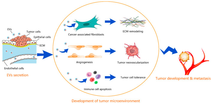
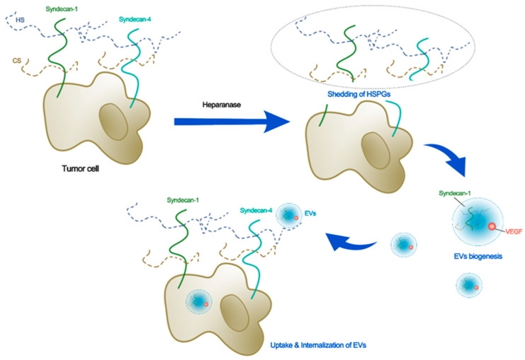
Angiogenesis in tumor growth and progression involves a series of complex changes in the tumor microenvironment. Extracellular vesicles (EVs) are important components of the tumor microenvironment, which can be classified as exosomes, apoptotic vesicles, and matrix vesicles according to their origins and properties. The EVs that share many common biological properties are important factors for the microenvironmental modification and play a vital role in tumor growth and progression. For example, vascular endothelial growth factor (VEGF) exosomes, which carry VEGF, participate in the tolerance of anti-angiogenic therapy (AAT). The glycocalyx is a mucopolysaccharide structure consisting of glycoproteins, proteoglycans, and glycosaminoglycans. Both endothelial and tumor cells have glycocalyx at their surfaces. Glycocalyx at both cells mediates the secretion and uptake of EVs. On the other hand, many components carried by EVs can modify the glycocalyx, which finally facilitates the development of the tumor microenvironment. In this short review, we first summarize the role of EVs in the development of the tumor microenvironment. Then we review how the glycocalyx is associated with the tumor microenvironment and how it is modulated by the EVs, and finally, we review the role of the glycocalyx in the synthesis, release, and uptake of EVs that affect tumor microenvironments. This review aims to provide a basis for the mechanistic study of AAT and new clues to address the challenges in AAT tolerance, tumor angiogenesis and metastasis.
Keywords: angiogenesis; extracellular vesicles; glycocalyx; metastasis; microenvironment.
Conflict of interest statement
The authors declare no conflict of interest.
Figures
References
-
- Cacheux W., Boisserie T., Staudacher L., Vignaux O., Dousset B., Soubrane O., Terris B., Mateus C., Chaussade S., Goldwasser F. Reversible tumor growth acceleration following bevacizumab interruption in metastatic colorectal cancer patients scheduled for surgery. Ann. Oncol. Off. J. Eur. Soc. Med. Oncol. 2008;19:1659–1661. doi: 10.1093/annonc/mdn540. - DOI - PubMed
-
- Becherirat S., Valamanesh F., Karimi M., Faussat A.M., Launay J.M., Pimpie C., Therwath A., Pocard M. Discontinuous Schedule of Bevacizumab in Colorectal Cancer Induces Accelerated Tumor Growth and Phenotypic Changes. Transl. Oncol. 2018;11:406–415. doi: 10.1016/j.tranon.2018.01.017. - DOI - PMC - PubMed
-
- Griffioen A.W., Mans L.A., de Graaf A.M.A., Nowak-Sliwinska P., de Hoog C., de Jong T.A.M., Vyth-Dreese F.A., van Beijnum J.R., Bex A., Jonasch E. Rapid angiogenesis onset after discontinuation of sunitinib treatment of renal cell carcinoma patients. Clin. Cancer Res. Off. J. Am. Assoc. Cancer Res. 2012;18:3961–3971. doi: 10.1158/1078-0432.CCR-12-0002. - DOI - PMC - PubMed
Publication types
Grants and funding
LinkOut - more resources
Full Text Sources
Research Materials
Miscellaneous
