. 2022 Nov 5;14(21):5447.
doi: 10.3390/cancers14215447.

Testicular Neoplasms: Primary Tumour Size Is Closely Interrelated with Histology, Clinical Staging, and Tumour Marker Expression Rates-A Comprehensive Statistical Analysis

Affiliations

Testicular Neoplasms: Primary Tumour Size Is Closely Interrelated with Histology, Clinical Staging, and Tumour Marker Expression Rates-A Comprehensive Statistical Analysis

Klaus-Peter Dieckmann et al. Cancers (Basel). .

Abstract

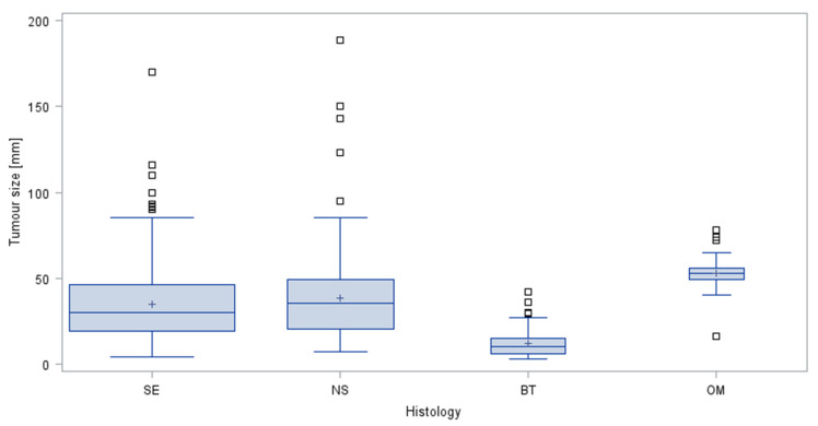
The role of primary tumour size (TS) in the clinical course of testicular tumours is incompletely understood. We retrospectively evaluated 641 consecutive patients with testicular neoplasms with regard to TS, histology, clinical stage (CS), serum tumour marker (STM) expression and patient age using descriptive statistical methods. TS ≤ 10 mm was encountered in 13.6% of cases. Median TS of 10 mm, 30 mm, 35 mm, and 53 mm were found in benign tumours, seminomas, nonseminomas, and other malignant tumours, respectively. In cases with TS ≤ 10 mm, 50.6% had benign tumours. Upon receiver operating characteristics analysis, TS of > 16 mm revealed 81.5% sensitivity and 81.0% specificity for detecting malignancy. In subcentimeter germ cell tumours (GCTs), 97.7% of cases had CS1, and CS1 frequency dropped with increasing TS. Expression rates of all STMs significantly increased with TS. MicroRNA-371a-3p (M371) serum levels had higher expression rates than classical STMs, with a rate of 44.1% in subcentimeter GCTs. In all, TS is a biologically relevant factor owing to its significant associations with CS, STM expression rates and histology. Importantly, 50% of subcentimeter testicular neoplasms are of benign nature, and M371 outperforms the classical markers even in subcentimeter tumours.

Keywords: alpha-fetoprotein; germ cell tumour; human chorionic gonadotropin; microRNA; seminoma; testicular tumour; tumour marker; tumour size.


Conflict of interest statement

K-PD and GB declare ownership of shares, each of 8.9%, in mirdetect GmbH, Bremerhaven, Germany. The remaining authors declare that the research was conducted in the absence of any commercial or financial relationships that could be construed as a potential conflict of interest.

Figures

Figure 1
Figure 1
Tumour sizes observed in the four histologic groups. Box and whisker plots showing the distribution of tumour sizes stratified by histologic subtypes of testicular neoplasms. The boxes display the first quartile, median and third quartile. The whiskers are defined as the largest or lowest observed value that falls within 1.5 times the interquartile range measured from Q3 or Q1, respectively. Area of box relates to sample size. □ outliers; + denotes arithmetic mean; SE seminoma; NS nonseminoma; BT benign tumours; OM other malignancies.
Figure 2
Figure 2
Typical subcentimeter testicular neoplasm. Surgical specimen of a 6 mm sized benign Leydig cell tumour excised by testis sparing surgery.
Figure 3
Figure 3
Proportions of histologic subgroups in testicular tumours sized >10 mm and ≤10 mm.
Figure 4
Figure 4
ROC analysis for predicting histology of a testicular neoplasm by its size.
Figure 5
Figure 5
Probability curve for prediction of malignant histology of a testicular neoplasm. The logistic regression curve indicates the probability of a given tumour size to predict malignancy. Shadowed area represents 95% confidence intervals. Neoplasms with a size of ≤8 mm involve a 50% probability of malignancy, while tumour sizes of ≥25 mm, ≥33 mm, and ≥39 mm involve probabilities of 95%, 99%, and 100%, respectively.
Figure 6
Figure 6
Tumour sizes in clinical stages in germ cell tumours. Box and whisker plots showing the distribution of tumour sizes stratified by clinical stages of testicular neoplasms. The boxes display the first quartile, median and third quartile. The whiskers are defined as the largest or lowest observed value that falls within 1.5 times the interquartile range measured from Q3 or Q1, respectively. Area of box denotes sample size. □ outliers; + denotes arithmetic mean; CS clinical stage.
Figure 7
Figure 7
Expression rates of M371 and AFP and/or bHCG in germ cell tumours in relation to size of primary tumour in seminoma (A) and in nonseminoma (B). Blue columns denote expression rates of AFP and/or bHCG in five categories of tumour size, red columns indicate expression rates of M371. Overall, the expression rates of all tumour markers were higher in nonseminoma than in seminoma. All markers showed a significant trend towards lower expression rates with decreasing tumour size. M371 had higher expression rates than the other markers even in the smallest tumour size category. Error bars represent 95% CIs.

Comment in

References

    1. Boden G., Gibb R. Radiotherapy and testicular neoplasms. Lancet. 1951;2:1195–1197. doi: 10.1016/S0140-6736(51)93202-3. - DOI - PubMed
    1. Dixon F.J., Moore R.A. Testicular tumors. A Clinicopathologic study. Cancer. 1953;6:427–454. doi: 10.1002/1097-0142(195305)6:3<427::AID-CNCR2820060302>3.0.CO;2-U. - DOI - PubMed
    1. Javadpour N. The National Cancer Institute Experience with Testicular Cancer. J. Urol. 1978;120:651–659. doi: 10.1016/S0022-5347(17)57316-4. - DOI - PubMed
    1. Collins D.H., Pugh R.C.B. Classification and frequency of testicular tumours. Br. J. Urol. 1964;36:1–11. - PubMed
    1. Fraley E.E., Lange P.H., Kennedy B.J. Germ-cell testicular cancer in adults (first of two parts) N. Engl. J. Med. 1979;301:1370–1377. doi: 10.1056/NEJM197912203012504. - DOI - PubMed

LinkOut - more resources