

Intravascular NK/T-Cell Lymphoma: What We Know about This Diagnostically Challenging, Aggressive Disease
- PMID: 36358876
- PMCID: PMC9658079
- DOI: 10.3390/cancers14215458
Intravascular NK/T-Cell Lymphoma: What We Know about This Diagnostically Challenging, Aggressive Disease
Abstract
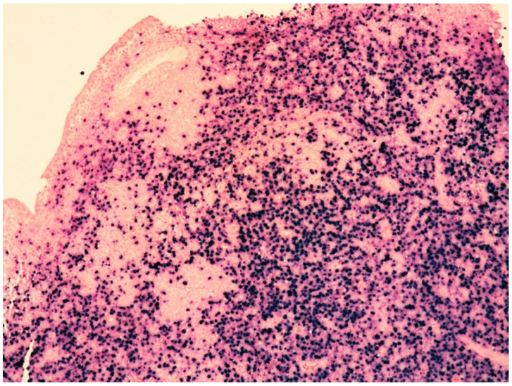
Intravascular lymphoma is a form of lymphoid malignancy characterized by neoplastic cells growing almost exclusively within the lumina of small- to medium-sized blood vessels. Most cases are of B-cell origin with rare cases of natural killer or T-cell lineage. Extranodal sites are affected, mainly the skin and central nervous system, although any organ may be involved. Intravascular NK/T-cell lymphoma deserves special attention because of its clinicopathologic features and the need for adequate immunophenotyping combined with clonality test for a proper diagnosis. Moreover, intravascular NK/T-cell lymphoma is strongly linked to Epstein-Barr virus (EBV), which is considered to play a role in tumorigenesis and to be responsible for the aggressive behavior of the disease. In this paper, we review the current knowledge on this rare lymphoma and, in particular, the most recent advances about its molecular landscape. The main distinguishing features with other EBV-related entities, such as extranodal NK/T-cell lymphoma, EBV-positive primary nodal T/NK-cell lymphoma, and aggressive NK-cell leukemia, are discussed to help pathologists obtain the correct diagnosis and consequently develop an adequate and prompt therapy response.
Keywords: EBV-positive primary nodal T/NK-cell lymphoma; Epstein–Barr virus; aggressive NK-cell leukemia; extranodal NK/T-cell lymphoma; intravascular NK/T-cell lymphoma.
Conflict of interest statement
The authors declare no conflict of interest.
Figures
Similar articles
-
Intravascular NK/T-cell lymphoma: a case report and literature review.J Pathol Transl Med. 2023 Nov;57(6):332-336. doi: 10.4132/jptm.2023.10.30. Epub 2023 Nov 14. J Pathol Transl Med. 2023. PMID: 37981727 Free PMC article.
-
Epstein-Barr virus-associated T- and NK-cell lymphoproliferative diseases: an update and diagnostic approach.Pathology. 2020 Jan;52(1):111-127. doi: 10.1016/j.pathol.2019.09.011. Epub 2019 Nov 22. Pathology. 2020. PMID: 31767131 Review.
-
Nonnasal lymphoma expressing the natural killer cell marker CD56: a clinicopathologic study of 49 cases of an uncommon aggressive neoplasm.Blood. 1997 Jun 15;89(12):4501-13. Blood. 1997. PMID: 9192774 Review.
-
Intravascular large T-cell or NK-cell lymphoma: a rare variant of intravascular large cell lymphoma with frequent cytotoxic phenotype and association with Epstein-Barr virus infection.Am J Surg Pathol. 2008 Jun;32(6):891-8. doi: 10.1097/PAS.0b013e31815d29c9. Am J Surg Pathol. 2008. PMID: 18425045
-
Intravascular NK/T-cell lymphoma: a report of five cases with cutaneous manifestation from China.J Cutan Pathol. 2015 Sep;42(9):610-7. doi: 10.1111/cup.12515. Epub 2015 May 29. J Cutan Pathol. 2015. PMID: 25931234
Cited by
-
Cutaneous Intravascular Hematolymphoid Entities: A Review.Diagnostics (Basel). 2024 Mar 23;14(7):679. doi: 10.3390/diagnostics14070679. Diagnostics (Basel). 2024. PMID: 38611591 Free PMC article. Review.
-
Intravascular NK/T-cell lymphoma: a case report and literature review.J Pathol Transl Med. 2023 Nov;57(6):332-336. doi: 10.4132/jptm.2023.10.30. Epub 2023 Nov 14. J Pathol Transl Med. 2023. PMID: 37981727 Free PMC article.
-
[Chinese expert consensus on the diagnosis and management of intravascular large B cell lymphoma (2023)].Zhonghua Xue Ye Xue Za Zhi. 2023 Mar 14;44(3):177-181. doi: 10.3760/cma.j.issn.0253-2727.2023.03.001. Zhonghua Xue Ye Xue Za Zhi. 2023. PMID: 37356977 Free PMC article. Chinese. No abstract available.
-
Intravascular Lymphoma: A Unique Pattern Underlying a Protean Disease.Cancers (Basel). 2025 Jul 15;17(14):2355. doi: 10.3390/cancers17142355. Cancers (Basel). 2025. PMID: 40723240 Free PMC article. Review.
-
Molecular Insights into the Diagnosis of Anaplastic Large Cell Lymphoma: Beyond Morphology and Immunophenotype.Int J Mol Sci. 2025 Jun 19;26(12):5871. doi: 10.3390/ijms26125871. Int J Mol Sci. 2025. PMID: 40565334 Free PMC article. Review.
References
-
- Santucci M., Pimpinelli N., Massi D., Kadin M.E., Meijer C.J.L.M., Muller-Hermelink H.K., Paulli M., Wechsler J., Willemze R., Audrig H., et al. EORTC cutaneous Lymphoma Task Force. Cytotoxic/natural killer cell cutaneous lymphomas. Report of EORTC cutaneous lymphoma Task Force workshop. Cancer. 2003;97:610–627. doi: 10.1002/cncr.11107. - DOI - PubMed
Publication types
LinkOut - more resources
Full Text Sources
