. 2022 Oct 28;12(11):1584.
doi: 10.3390/biom12111584.

Submilligram Level of Beetle Antifreeze Proteins Minimize Cold-Induced Cell Swelling and Promote Cell Survival

Affiliations

Submilligram Level of Beetle Antifreeze Proteins Minimize Cold-Induced Cell Swelling and Promote Cell Survival

Keiko Omori et al. Biomolecules. .

Abstract

Hypothermic (cold) preservation is a limiting factor for successful cell and tissue transplantation where cell swelling (edema) usually develops, impairing cell function. University of Wisconsin (UW) solution, a standard cold preservation solution, contains effective components to suppress hypothermia-induced cell swelling. Antifreeze proteins (AFPs) found in many cold-adapted organisms can prevent cold injury of the organisms. Here, the effects of a beetle AFP from Dendroides canadensis (DAFP-1) on pancreatic β-cells preservation were first investigated. As low as 500 µg/mL, DAFP-1 significantly minimized INS-1 cell swelling and subsequent cell death during 4 °C preservation in UW solution for up to three days. However, such significant cytoprotection was not observed by an AFP from Tenebrio molitor (TmAFP), a structural homologue to DAFP-1 but lacking arginine, at the same levels. The cytoprotective effect of DAFP-1 was further validated with the primary β-cells in the isolated rat pancreatic islets in UW solution. The submilligram level supplement of DAFP-1 to UW solution significantly increased the islet mass recovery after three days of cold preservation followed by rewarming. The protective effects of DAFP-1 in UW solution were discussed at a molecular level. The results indicate the potential of DAFP-1 to enhance cell survival during extended cold preservation.

Keywords: antifreeze protein; cell swelling; post-hypothermic preservation survival.


Conflict of interest statement

The authors declare no conflict of interest. The funders had no role in the design of the study; in the collection, analyses, or interpretation of data; in the writing of the manuscript, or in the decision to publish the results.

Figures
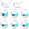
Figure 1
Figure 1
Survival of INS-1 cells cold preserved in UW solution supplemented with DAFP-1. INS-1 cells were cold preserved in UW solution alone, UW solution containing vehicle (PBS control), or with 250, 500, and 1000 µg/mL DAFP-1 for up to 72 h. (A) The live cell number and (B) the viability of INS-1 cells before and after the cold preservation in the UW solution or PBS control group tested at 24, 48, and 72 h of preservation. The data show the mean ± standard deviation of six independent experiments, * p < 0.05, ** p < 0.01, by the one-way ANOVA. The live cell number of INS-1 cells after (C) 24 h, (D) 48 h, and (E) 72 h of cold preservation was compared to pre-preservation shown by the live cell recovery (live cell number at each time point divided by the live cell number before preservation), and the INS-1 cell viability of each protein group was compared to the UW solution group at (F) 24 h, (G) 48 h, and (H) 72 h shown as viability index (viability of INS-1 cells preserved in each condition divided by the viability of INS-1 cells preserved in the UW solution). The data show the mean ± standard error of five to six independent experiments (open circle for UW alone, open square for PBS control, solid square, solid triangle, and solid circle for 250, 500 and 1000 µg/mL DAFP-1, respectively), * p < 0.05 and ** p < 0.01 by the one-way ANOVA.
Figure 2
Figure 2
Changes in the size of INS-1 cells cold preserved in the UW solution supplemented with DAFP-1. The INS-1 cells were cold preserved in UW alone, UW solution containing vehicle (PBS control) or with 250, 500, and 1000 µg/mL DAFP-1 for up to 72 h, and the live cell sizes were analyzed along with the cell viability using the trypan blue staining. The percent increases of the live cell size before- and post-cold preservation was calculated as the difference of the mean diameter of the live cells between the post-preservation and pre-preservation divided by that of the pre-preservation at (A) 24 h, (B) 48 h, and (C) 72 h after cold preservation (open circle for UW only, open square for PBS control, solid square, solid triangle and solid circle for DAFP-1 250, 500 and 1000 µg/mL, respectively). The data show the mean ± standard error of five to six independent experiments, * p < 0.05 by the one-way ANOVA. (D) The overall correlation between the percentage of cell size increase and the viability index of the corresponding samples from all the groups at 48 h preservation and (E) 72 h preservation (r = −0.57, r2 = 0.33, p < 0.005, N = 5 independent experiments with 29 data points).
Figure 3
Figure 3
Fragments of poly(ADP-ribose) polymerase-1 (PARP-1) in the cold-preserved INS-1 cells. The total length and cleaved PARP-1 protein expressions in the INS-1 cells before and after 72 h of cold preservation with or without 500 µg/mL DAFP-1 were assessed by Western blot. (A) Representative image of the Western blot bands for the total length PARP-1, cleaved PARP-1, and β-actin, (B) quantified relative protein expression levels for the cleaved PARP-1 over β-actin, and (C) cleaved PARP-1 over total length PARP-1 (open circle for pre preservation, open square for 0 µg/mL, and solid triangle for 500 µg/mL DAFP-1). The data show the mean ± standard error of five independent experiments, * p < 0.05 by the Student t-test.
Figure 4
Figure 4
Potency of DAFP-1 in maintaining cold-preserved INS-1 cell viability compared to TmAFP and BSA. The viability index of the cold-preserved INS-1 cells in UW solution supplemented with TmAFP at a different concentration (0, 250, 500, and 1000 µg/mL) for (A) 24 h and (B) 72 h. The cell viability of each protein condition was normalized by the viability of the UW solution group (AFP 0 µg/mL, open circle) at each time point (solid square, solid triangle, and solid circle for 250, 500, and 1000 µg/mL DAFP-1, respectively). (C) The dose-dependent changes of viability indexes for TmAFP and DAFP-1 at 72 h. The data show the mean ± standard error of five to six independent experiments, * p < 0.05 by the Student t-test.
Figure 5
Figure 5
Viability, survival, and function of cold-preserved rat islets. Isolated rat islets were cold preserved in UW solution at 4 °C with or without DAFP-1 (0, 250, or 500 µg/mL) for up to 72 h. (A) The changes in cold-preserved islet viability measured by the fluorescent diacetate (FDA) and propidium iodide (PI) staining, and (B) Viability Index of each protein condition (solid square for 250 µg/mL DAFP-1 and solid triangle for 500 µg/mL DAFP-1) calculated relative to the control (DAFP-1 0 µg/mL = UW solution alone, open circle). In the separate experiment, rat islets were cold preserved in UW solution with or without DAFP-1 (0, 250, or 500 µg/mL) for 72 h, followed by overnight rewarming in the culture at 27 °C. (C) The recovery index and (D) viability index of rewarmed islets was shown as relative to the control at each time point. (E) The representative images of the 72 h cold-preserved and rewarmed (72 h C + R) rat islet viability assessed with FDA (alive in green)/PI (dead in orange) staining. Scale bar: 200 µm. (F) Overall survival after the 72 h cold preservation followed by the rewarming was calculated by the recovery index multiplied by the viability (%) relative to the pre-preservation (solid circle). (G) The function of rat islets after 72 h of cold preservation followed by rewarming assessed by the glucose-stimulated insulin release in a static incubation assay (1 h incubation assay in 2.8 mM glucose followed by 1 h incubation in 28 mM glucose) and (H) stimulation index calculated by the insulin release during the 28 mM glucose incubation divided by the insulin release during the 2.8 mM glucose incubation. The data show the mean ± standard error of three to four independent experiments, * p < 0.05, *** p < 0.005 by the Student’s t-test.
Figure 6
Figure 6
Structures and a schematic diagram for the enhancement effect. (A) Structure of raffinose with carboxylate group in red, (B) structure of lactobionate, and (C) structural overlay of DAFP-1 (cyan) and TmAFP (yellow–orange). The arginine residues in DAFP-1 are shown in licorice representation and colored by element (C in cyan, N in blue, O in red, and H removed for clarity). The guanidinium group (shown in blue) of an arginine residue is represented in a large aside of the protein’s structure. (D) Schematic representation of DAFP-1 enhancing the function of cell membrane impermeant compounds in UW solution, such as lactobionate (represented as negatively charged hexagons in orange) and raffinose (represented as decagons in blue).

Similar articles

References

    1. Southard J.H., van Gulik T.M., Ametani M.S., Vreugdenhil P.K., Lindell S.L., Pienaar B.L., Belzer F.O. Important components of the UW solution. Transplantation. 1990;49:251–257. doi: 10.1097/00007890-199002000-00004. - DOI - PubMed
    1. Wahlberg J.A., Southard J.H., Belzer F.O. Development of a cold storage solution for pancreas preservation. Cryobiology. 1986;23:477–482. doi: 10.1016/0011-2240(86)90056-8. - DOI - PubMed
    1. Squifflet J.P., LeDinh H., de Roover A., Meurisse M. Pancreas Preservation for Pancreas and Islet Transplantation: A Minireview. Transplant. Proc. 2011;43:3398–3401. doi: 10.1016/j.transproceed.2011.09.052. - DOI - PubMed
    1. Raymond J.A., DeVries A.L. Adsorption Inhibition as a Mechanism of Freezing Resistance in Polar Fishes. Proc. Natl. Acad. Sci. USA. 1977;74:2589–2593. doi: 10.1073/pnas.74.6.2589. - DOI - PMC - PubMed
    1. Bhatnagar B., Zakharov B., Fisyuk A., Wen X., Karim F., Lee K., Seryotkin Y., Mogodi M., Fitch A., Boldyreva E., et al. Protein/Ice Interaction: High-Resolution Synchrotron X-ray Diffraction Differentiates Pharmaceutical Proteins from Lysozyme. J. Phys. Chem. B. 2019;123:5690–5699. doi: 10.1021/acs.jpcb.9b02443. - DOI - PMC - PubMed

Publication types