

Androgen Signaling in Uterine Diseases: New Insights and New Targets
- PMID: 36358974
- PMCID: PMC9687413
- DOI: 10.3390/biom12111624
Androgen Signaling in Uterine Diseases: New Insights and New Targets
Abstract
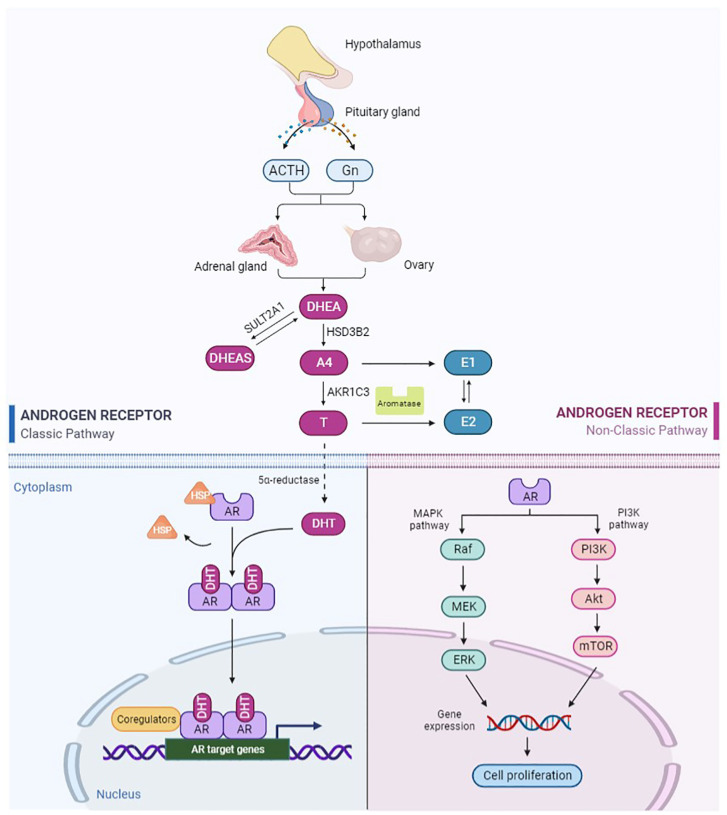
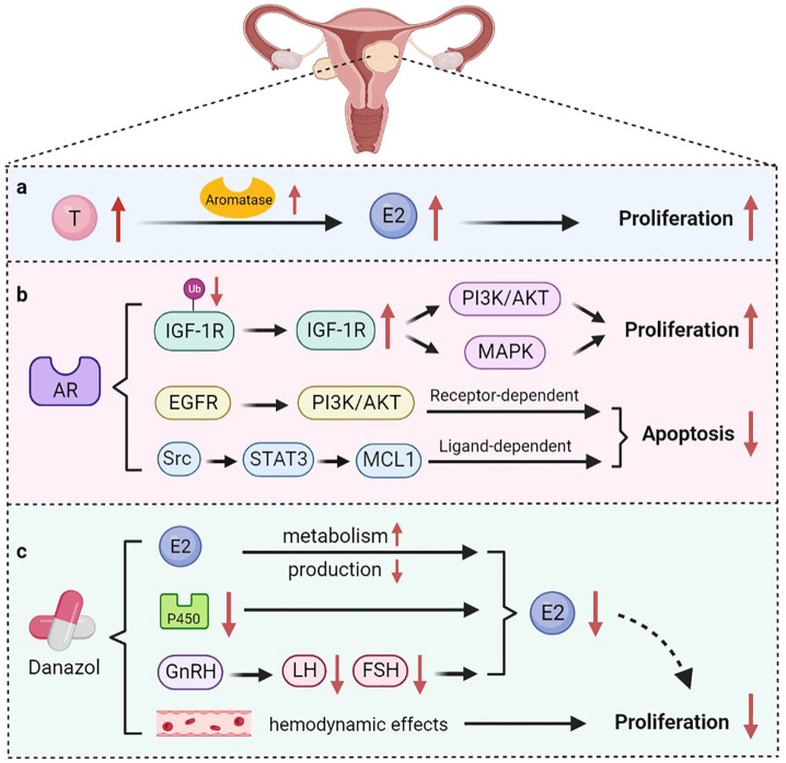
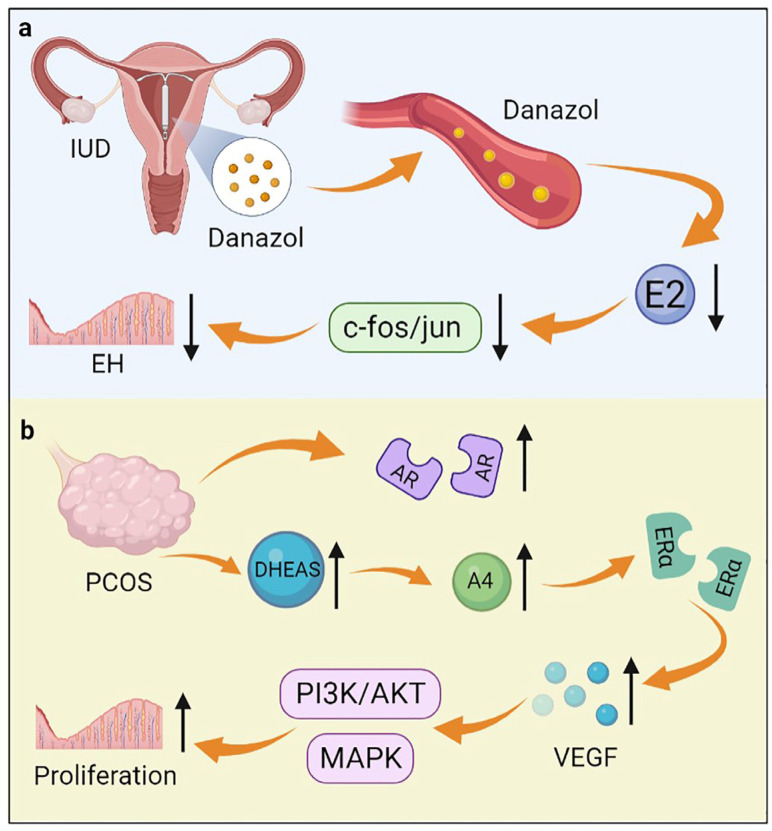
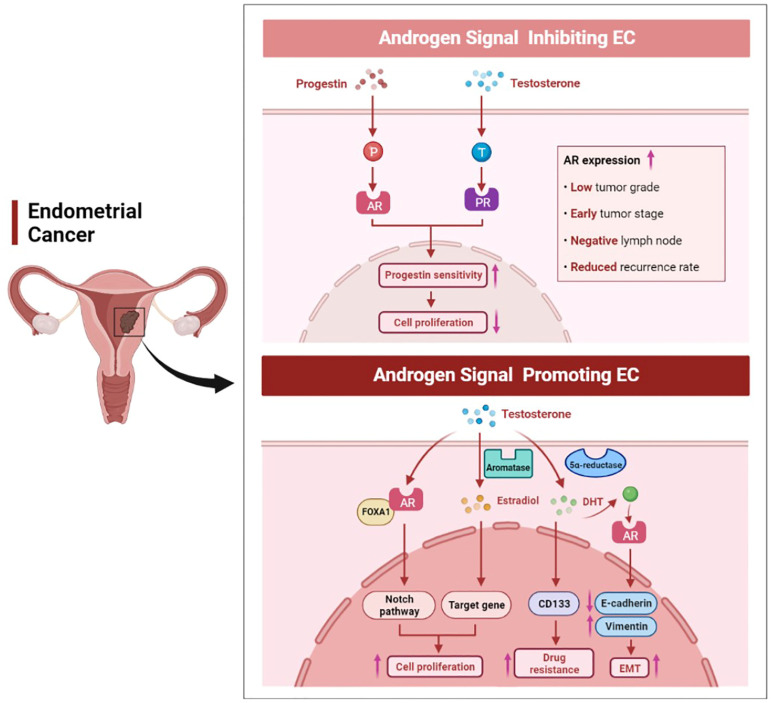
Common uterine diseases include endometriosis, uterine fibroids, endometrial polyps, endometrial hyperplasia, endometrial cancer, and endometrial dysfunction causing infertility. Patients with uterine diseases often suffer from abdominal pain, menorrhagia, infertility and other symptoms, which seriously impair their health and disturb their lives. Androgens play important roles in the normal physiological functions of the uterus and pathological progress of uterine diseases. Androgens in women are synthesized in the ovaries and adrenal glands. The action of androgens in the uterus is mainly mediated by its ligand androgen receptor (AR) that regulates transcription of the target genes. However, much less is known about the signaling pathways through which androgen functions in uterine diseases, and contradictory findings have been reported. This review summarizes and discusses the progress of research on androgens and the involvement of AR in uterine diseases. Future studies should focus on developing new therapeutic strategies that precisely target specific AR and their related signaling pathways in uterine diseases.
Keywords: androgen; androgen receptor; endometrium; uterine diseases.
Conflict of interest statement
The authors declare no conflict of interest.
Figures
References
-
- Allen N.E., Key T.J., Dossus L., Rinaldi S., Cust A., Lukanova A., Peeters P.H., Onland-Moret N.C., Lahmann P.H., Berrino F., et al. Endogenous sex hormones and endometrial cancer risk in women in the European Prospective Investigation into Cancer and Nutrition (EPIC) Endocr. -Relat. Cancer. 2008;15:485–497. doi: 10.1677/ERC-07-0064. - DOI - PMC - PubMed
-
- Labrie F., Simard J., Luu-The V., Bélanger A., Pelletier G. Structure, function and tissue-specific gene expression of 3β-hydroxysteroid dehydrogenase/5-ene-4-ene isomerase enzymes in classical and peripheral intracrine steroidogenic tissues. J. Steroid Biochem. Mol. Biol. 1992;43:805–826. doi: 10.1016/0960-0760(92)90308-6. - DOI - PubMed
Publication types
MeSH terms
Substances
LinkOut - more resources
Full Text Sources
Medical
Research Materials
