. 2022 Nov 4;12(11):1635.
doi: 10.3390/biom12111635.

Neuronal Firing and Glutamatergic Synapses in the Substantia Nigra Pars Reticulata of LRRK2-G2019S Mice

Affiliations

Neuronal Firing and Glutamatergic Synapses in the Substantia Nigra Pars Reticulata of LRRK2-G2019S Mice

Giacomo Sitzia et al. Biomolecules. .

Abstract

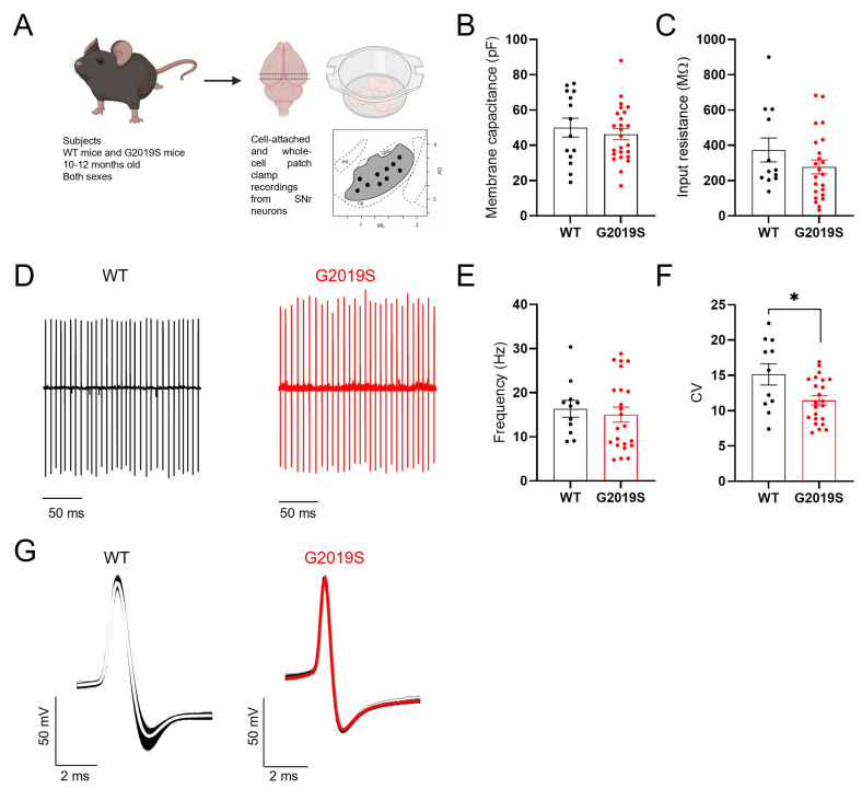
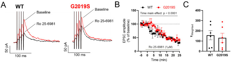
Pathogenic mutations in the leucine-rich repeat kinase 2 (LRRK2) gene are frequent causes of familial Parkinson's Disease (PD), an increasingly prevalent neurodegenerative disease that affects basal ganglia circuitry. The cellular effects of the G2019S mutation in the LRRK2 gene, the most common pathological mutation, have not been thoroughly investigated. In this study we used middle-aged mice carrying the LRRK2-G2019S mutation (G2019S mice) to identify potential alterations in the neurophysiological properties and characteristics of glutamatergic synaptic transmission in basal ganglia output neurons, i.e., substantia nigra pars reticulata (SNr) GABAergic neurons. We found that the intrinsic membrane properties and action potential properties were unaltered in G2019S mice compared to wild-type (WT) mice. The spontaneous firing frequency was similar, but we observed an increased regularity in the firing of SNr neurons recorded from G2019S mice. We examined the short-term plasticity of glutamatergic synaptic transmission, and we found an increased paired-pulse depression in G2019S mice compared to WT mice, indicating an increased probability of glutamate release in SNr neurons from G2019S mice. We measured synaptic transmission mediated by NMDA receptors and we found that the kinetics of synaptic responses mediated by these receptors were unaltered, as well as the contribution of the GluN2B subunit to these responses, in SNr neurons of G2019S mice compared to WT mice. These results demonstrate an overall maintenance of basic neurophysiological and synaptic characteristics, and subtle changes in the firing pattern and in glutamatergic synaptic transmission in basal ganglia output neurons that precede neurodegeneration of dopaminergic neurons in the LRRK2-G2019S mouse model of late-onset PD.

Keywords: LRRK2-G2019S; NMDA receptors; Parkinson’s disease; electrophysiology; glutamatergic synaptic transmission; substantia nigra reticulata.


Conflict of interest statement

The authors declare no conflict of interest. The funders had no role in the design of the study; in the collection, analyses, or interpretation of data; in the writing of the manuscript, or in the decision to publish the results.

Figures

Figure 1
Figure 1
Intrinsic properties of SNr neurons. (A) Scheme illustrating the experimental model and the procedures. (B,C) No differences were found in the membrane capacitance (B) and input resistance (C) of SNr neurons between G2019S and WT mice (WT: n = 14 cells from N = 4 mice; G2019S: n = 25 cells from N = 9 mice). (D) Example traces of cell-attached recordings of the spontaneous AP firing in SNr neurons from a WT mouse and a G2019S mouse. (E) The firing frequency of SNr neurons was similar in G2019S mice and WT mice (WT: 16.32 ± 1.9 Hz, n = 11 cells from N = 5 mice; G2019S: 15.02 ± 1.7 Hz, n = 23 cells from N = 6 mice; Mann-Whitney test p = 0.3827). (F) The coefficient of variation of the interspike intervals (CV) was significantly smaller in SNr neurons from G2019S mice compared to WT mice (WT: 15.0 ± 1.51, n = 11 cells from N = 5 mice; G2019S: 11.46 ± 0.65, n = 23 cells from N = 6 mice; unpaired t-test * p = 0.0130), indicating a more regular firing pattern. (G) Example traces of action potentials, measured in whole-cell current-clamp mode, in SNr neurons from a WT mouse and a G2019S mouse (see Table 1).
Figure 2
Figure 2
Glutamatergic synaptic transmission in SNr neurons. (A) Example traces of whole-cell voltage-clamp recordings of sEPSCs obtained in two SNr neurons from a WT mouse and a G2019S mouse. (B) The amplitude of sEPSCs recorded from SNr neurons was not different between WT and G2019S mice (WT: 15.09 ± 2.1 pA, n = 12 cells from N = 6 mice; G2019S: 15.74 ± 2.3 pA, n = 12 cells from N = 5 mice; Mann-Whitney test, p = 0.71). (C) The frequency of sEPSCs recorded from SNr neurons was not different between WT and G2019S mice (WT: 1.11 ± 0.23 Hz, n = 12 cells from N = 6 mice; G2019S: 0.89 ± 0.21 Hz, n = 12 cells from N = 5 mice; Mann-Whitney test, p = 0.4). (D,E) The paired-pulse ratio (PPR) of paired EPSCs evoked in SNr neurons at different stimulus intervals showed a significant increase in paired-pulse depression in G2019S mice when compared to WT mice (WT: n = 11 cells from N = 8 mice; G2019S: n = 10 cells from N = 9 mice; repeated measures two-way ANOVA, factors genotype and interval, main effect of genotype: F1, 19 = 4.405, * p = 0.0494).
Figure 3
Figure 3
Unaltered synaptic NMDARs in SNr neurons of G2019S. (A) Example traces of NMDAR-EPSCs—before (red traces) and during (black traces) bath application of the GluN2B antagonist Ro 25-691 (1 µM)—recorded in two SNr neurons from a WT mouse and a G2019S mouse. (B) Time course of the effect of Ro 25-2981, applied at the time indicated by the horizontal bar, on NMDAR-EPSC amplitude in SNr neurons from WT and G2019S mice. Ro 25-691 inhibited the NMDAR-EPSC to a similar level in WT and G2019S mice (WT: n = 3 cells from N = 3 mice; G2019S: n = 4 cells from N = 4 mice; 2-way ANOVA, factors genotype and time, main effect of time: F35, 188 = 11.61, p < 0.0001). (C) No differences were detected in the kinetic properties of NMDAR-EPSCs in the SNr between WT and G2019S mice (WT: n = 6 cells from N = 5 mice; G2019S: n = 6 cells from N = 5 mice).

References

    1. Gonzalez-Rodriguez P., Zampese E., Surmeier D.J. Selective neuronal vulnerability in Parkinson’s disease. Prog. Brain Res. 2020;252:61–89. - PubMed
    1. Pirkevi C., Lesage S., Brice A., Basak A.N. From genes to proteins in mendelian Parkinson’s disease: An overview. Anat. Rec. 2009;292:1893–1901. doi: 10.1002/ar.20968. - DOI - PubMed
    1. Yue M., Hinkle K.M., Davies P., Trushina E., Fiesel F.C., Christenson T.A., Schroeder A.S., Zhang L., Bowles E., Behrouz B., et al. Progressive dopaminergic alterations and mitochondrial abnormalities in LRRK2 G2019S knock-in mice. Neurobiol. Dis. 2015;78:172–195. doi: 10.1016/j.nbd.2015.02.031. - DOI - PMC - PubMed
    1. Healy D.G., Falchi M., O’Sullivan S.S., Bonifati V., Durr A., Bressman S., Brice A., Aasly J., Zabetian C.P., Goldwurm S., et al. Phenotype, genotype, and worldwide genetic penetrance of LRRK2-associated Parkinson’s disease: A case-control study. Lancet Neurol. 2008;7:583–590. doi: 10.1016/S1474-4422(08)70117-0. - DOI - PMC - PubMed
    1. Di Maio R., Hoffman E.K., Rocha E.M., Keeney M.T., Sanders L.H., De Miranda B.R., Zharikov A., Van Laar A., Stepan A.F. LRRK2 activation in idiopathic Parkinson’s disease. Sci. Transl. Med. 2018;10:eaar5429. doi: 10.1126/scitranslmed.aar5429. - DOI - PMC - PubMed

Publication types

MeSH terms

Substances