. 2022 Oct 28;12(21):2966.
doi: 10.3390/ani12212966.

Equine Stomach Development in the Fetal Period: An Anatomical, Topographical, and Morphometric Study

Affiliations

Equine Stomach Development in the Fetal Period: An Anatomical, Topographical, and Morphometric Study

Dominik Poradowski et al. Animals (Basel). .

Abstract

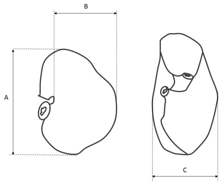
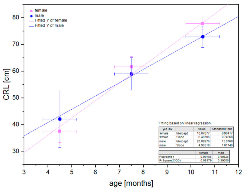
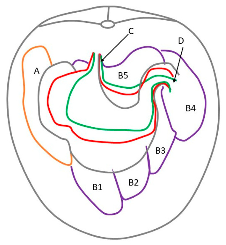
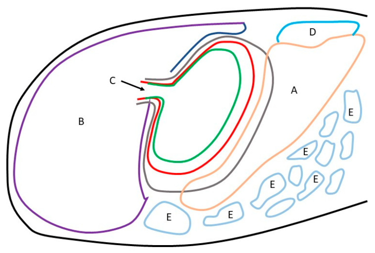
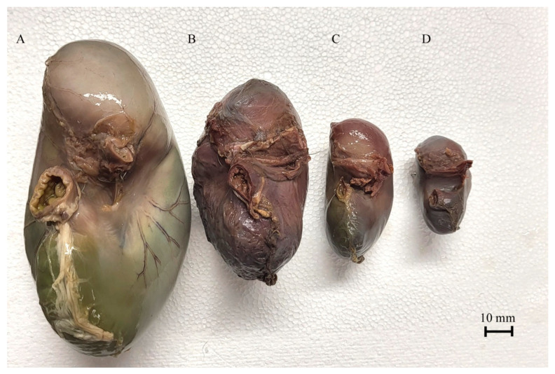
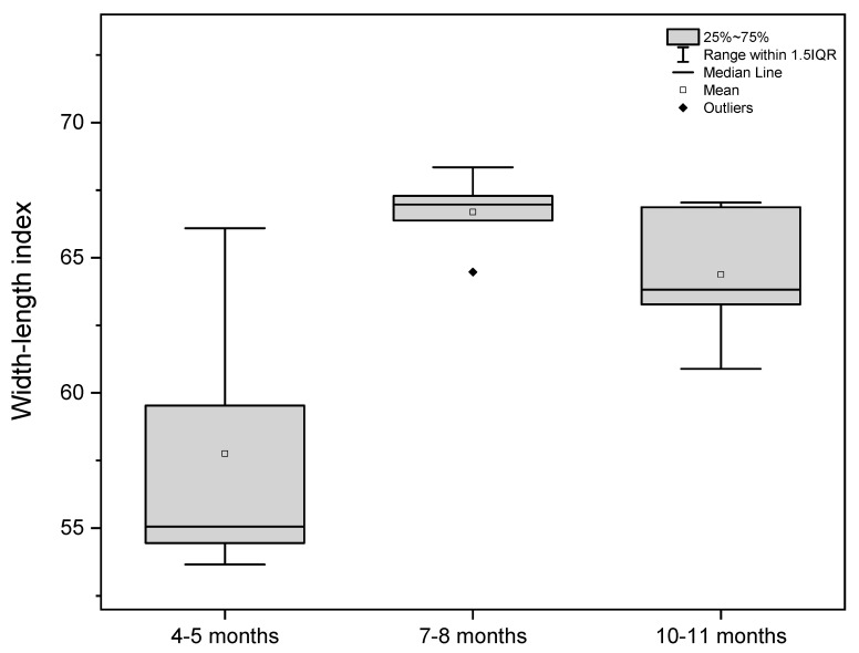
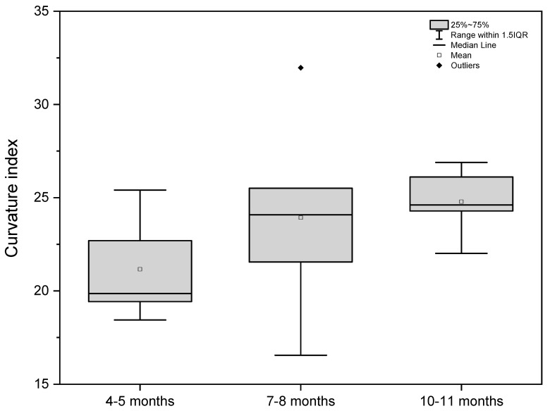
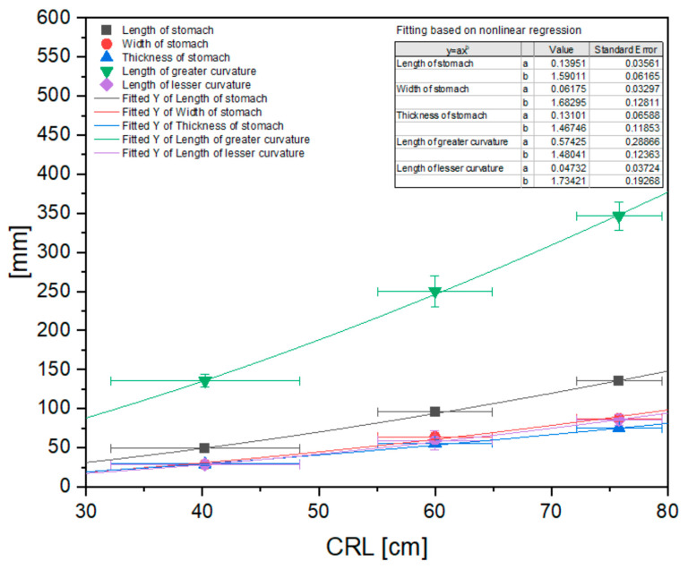
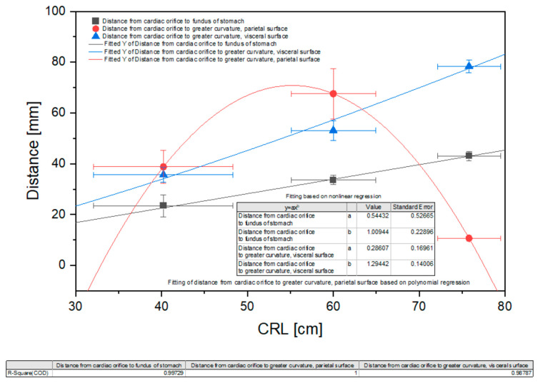
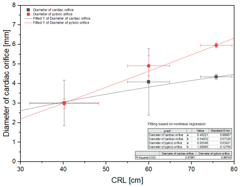
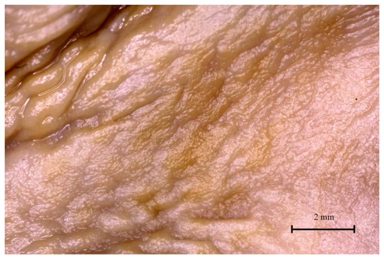
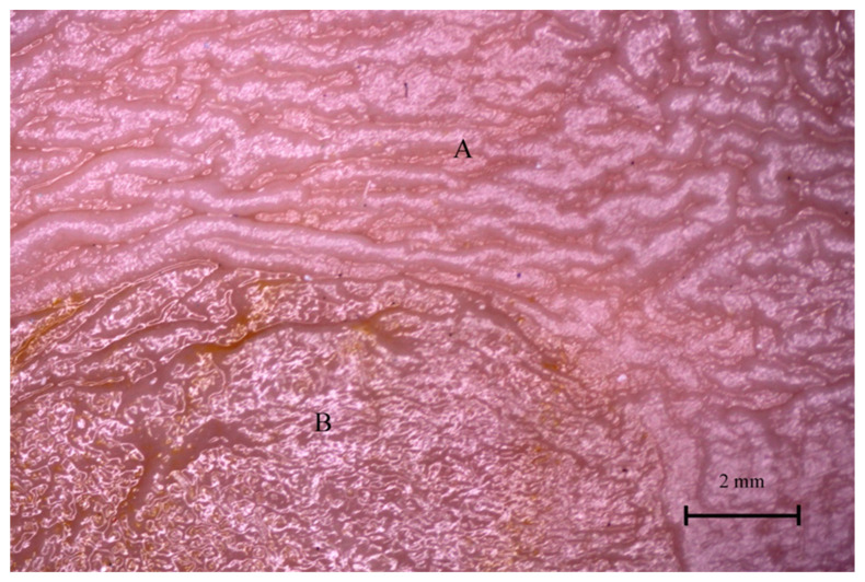
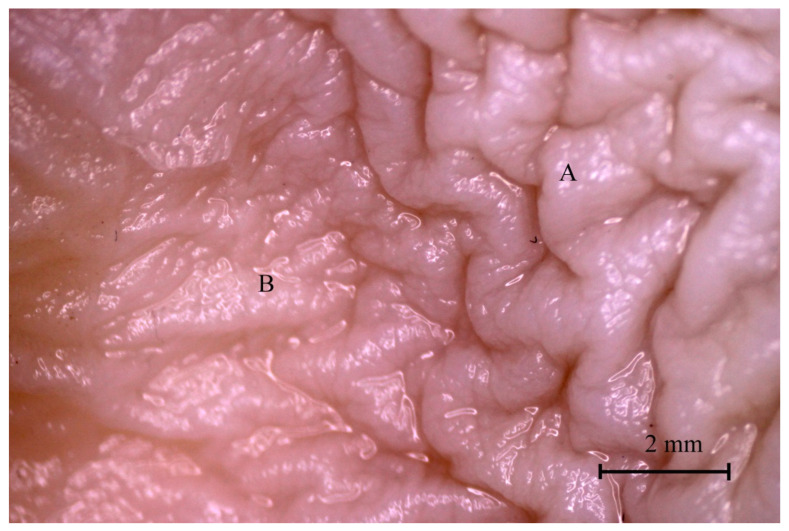
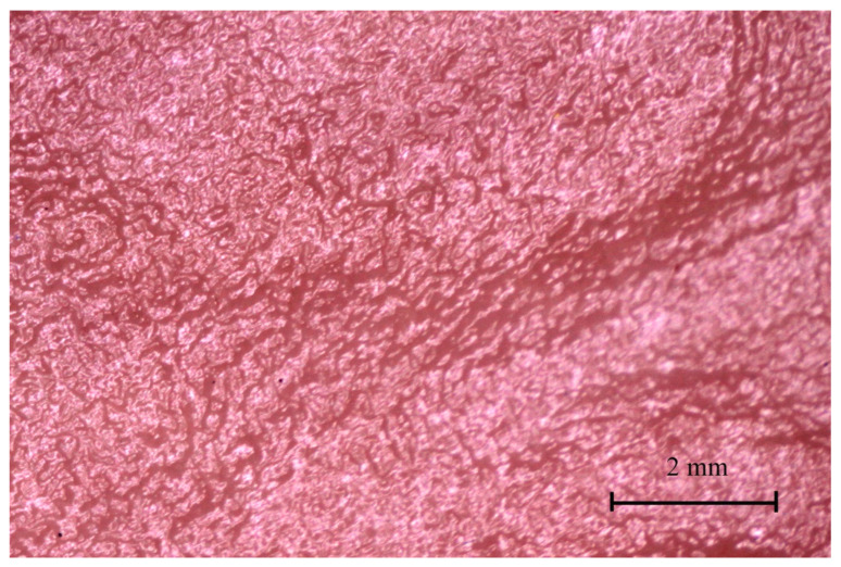
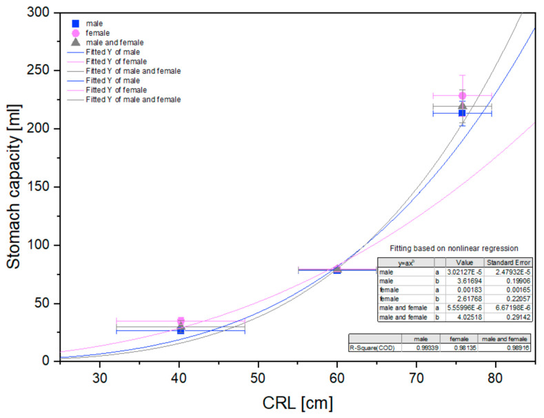
Studies of equine stomach prenatal development are very rare, and descriptions usually focus on the processes taking place in the embryonic period. Only general information about gastric organogenesis in the fetal period is available in embryology textbooks on domestic mammals. The material for our study included twenty half-breed horse fetuses divided into three age groups on the basis of known fetal age (verified using the CRL method). Our study consists of the topographical, morphological, and morphometrical description of stomach development between the 4th and 11th months of gestation. Even though the skeletotopy, syntopy, and holotopy of the stomach in the fetal period seems to be relatively unchanged, the organ shape and the proportions between its anatomical parts differed in fetuses from the three age groups. The achieved results were statistically elaborated to estimate the dynamics of the stomach shape. This can be described as changing from medium-wide to wide and from slightly bent to sharply bent. A nonlinear correlation of all metric values with CRL in all age groups was observed. A positive allometric growth rate of different intensity was seen in all metric parameters. All the values increased as the fetal period progressed. Only the parietal surface growth rate gradually changed from strongly positive allometric in the first age group to strongly negative allometric in the third age group. A difference between the non-glandular and glandular mucosa of the stomach was visible in the first group. Development of a well-distinguishable plicated edge margin began in the second age group together with gastric pits and gastric areas. The third age group showed a well-developed gastric groove and angular incisura.

Keywords: anatomy; development; equine; fetal period; morphometry; stomach; topography.


Conflict of interest statement

Neither of the authors has any financial or personal relationships that could inappropriately influence or bias the content of the paper.

Figures

Figure 1
Figure 1
The scheme of the stomach morphometry. A—the length of the stomach; B—the width of the stomach; C—the thickness of the stomach.
Figure 2
Figure 2
Linear regression in CRL values in the fetal period for male and female horse fetuses.
Figure 3
Figure 3
Topography of the stomach in the fetal period (caudal view). Green—first age group; red—second age group; black—third age group; A—spleen; B1—liver, left lateral lobe; B2—liver, left medial lobe; B3—liver, quadrate lobe; B4—liver, right lobe; B5—liver, caudate lobe; C—cardiac orifice; D—pyloric orifice.
Figure 4
Figure 4
Topography of the stomach in the fetal period (lateral view). Green—first age group; red—second age group; black—third age group; A—spleen; B—liver; C—cardiac orifice; D—kidney; E—small and large intestine.
Figure 5
Figure 5
Equine fetal stomach (the lesser curvature view). A—the third age group, B—the second age group, C and D—the first age group.
Figure 6
Figure 6
Equine fetal stomach (the visceral surface view). A—the third age group, B—the second age group, C and D—the first age group.
Figure 7
Figure 7
Width-length index values in the fetal period (4th to 11th month of gestation).
Figure 8
Figure 8
Curvature index values in the fetal period (4th to 11th month of gestation).
Figure 9
Figure 9
Nonlinear regression of basic stomach morphometric values (length, width, and thickness, and length of the greater and lesser curvature). CRL—crown–rump length.
Figure 10
Figure 10
Nonlinear regression of the stomach morphometric values (distance from the cardiac orifice to the stomach fundus, from the cardiac orifice to the greater curvature and the parietal surface, and distance from the cardiac orifice to the greater curvature and the visceral surface). CRL—crown-rump length.
Figure 11
Figure 11
Nonlinear regression of the stomach morphometric values (diameter of the cardiac orifice and pyloric orifice). CRL—crown–rump length.
Figure 12
Figure 12
Gastric mucosa in the plicated edge margin region (the first age group).
Figure 13
Figure 13
Gastric mucosa in the plicated edge margin region (the second age group). A—non-glandular part, B—glandular part.
Figure 14
Figure 14
Gastric mucosa in the plicated edge margin region (the third age group). A—non-glandular part, B—glandular part.
Figure 15
Figure 15
Gastric mucosa of the stomach body (third age group).
Figure 16
Figure 16
Stomach volume in subsequent age groups in relation to CRL.

Similar articles

Cited by

References

    1. Bucca S., Fogarty U., Collins A., Small V. Assessment of feto-placental well-being in the mare from mid-gestation to term: Transrectal and transabdominal ultrasonographic features. Theriogenology. 2005;64:542–557. doi: 10.1016/j.theriogenology.2005.05.011. - DOI - PubMed
    1. Murase H., Endo Y., Tsuchiya T., Kotoyori Y., Shikichi M., Ito K., Sato F., Nambo Y. Ultrasonographic evaluation of equine fetal growth throughout gestation in normal mares using a convex transducer. J. Vet. Med. Sci. 2014;76:947–953. doi: 10.1292/jvms.13-0259. - DOI - PMC - PubMed
    1. Agnew M.E., Slack J., Stefanovski D., Linton J.K., Sertich P.L. Sonographic appearance of the late gestation equine fetal intestine. Theriogenology. 2019;138:121–126. doi: 10.1016/j.theriogenology.2019.07.006. - DOI - PubMed
    1. Lanci A., Castagnetti C., Ranciati S., Sergio C., Mariella J. A regression model including fetal orbit measurements to predict parturition in Standardbred mares with normal pregnancy. Theriogenology. 2019;126:153–158. doi: 10.1016/j.theriogenology.2018.12.020. - DOI - PubMed
    1. Bucca S., De Oliveira I.R.S., Cunanan J.C., Vinardell T., Troedsson M.H. Doppler indices of the equine fetal carotid artery throughout gestation. Theriogenology. 2020;156:196–204. doi: 10.1016/j.theriogenology.2020.07.009. - DOI - PubMed

LinkOut - more resources