. 2022 Oct 31;10(11):2765.
doi: 10.3390/biomedicines10112765.

Profibrotic Effects of Endothelin-1 on Fibroblasts Are Mediated by Aldosterone in Vitro: Relevance to the Pathogenesis and Therapy of Systemic Sclerosis and Pulmonary Arterial Hypertension

Affiliations

Profibrotic Effects of Endothelin-1 on Fibroblasts Are Mediated by Aldosterone in Vitro: Relevance to the Pathogenesis and Therapy of Systemic Sclerosis and Pulmonary Arterial Hypertension

Giuseppe Argentino et al. Biomedicines. .

Abstract

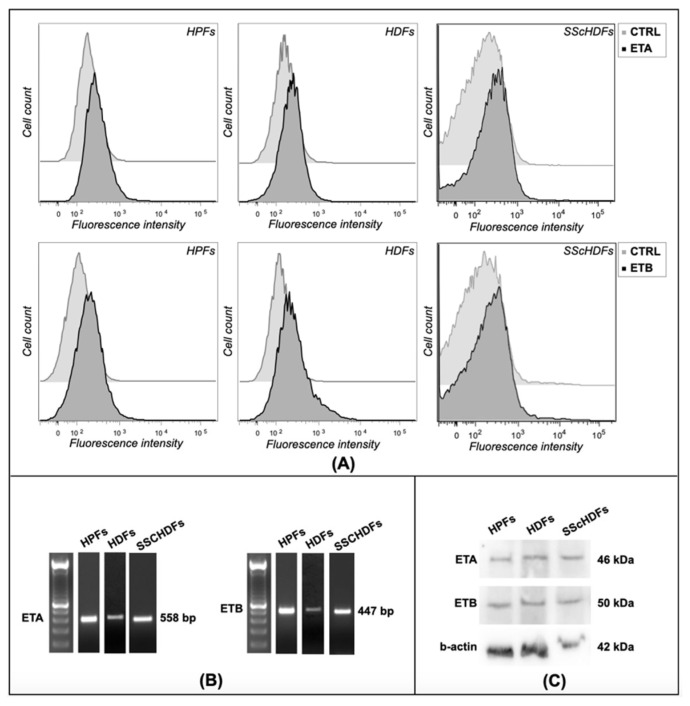
Endothelin-1 (ET-1) is a vasoactive and profibrotic peptide that plays a pivotal role in diseases such as systemic sclerosis (SSc) and pulmonary arterial hypertension (PAH), by inducing fibrosis and vascular remodeling. Such effects may be sustained by the induction of aldosterone production and reactive oxygen species (ROS). We have used fibroblasts obtained from skin of healthy donors and SSc patients and commercial fibroblasts from lung to evaluate whether ET-1 is able to stimulate ROS production directly or indirectly through aldosterone induction. We found that ET-1 receptors are present in all types of fibroblasts analyzed, whereas the expression of mineralocorticoid receptor (MCR) is lower in dermal fibroblasts from healthy donors (HDFs) compared to fibroblasts derived from lung (HPFs) or from skin of SSc patients (SScHDFs). ET-1 induces ROS production in HDFs and SScHDFs after 24 h of incubation involving its receptor B (ETB), whereas aldosterone exerts its effects after 40 min of incubation. Moreover, ROS production was inhibited by the pre-incubation of cells with MCR inhibitor. Our results indicate that ET-1 induces ROS indirectly through aldosterone production suggesting that aldosterone may play a pivotal role in the pathogenesis of SSc and PAH.

Keywords: aldosterone; endothelin-1; fibroblasts; pulmonary arterial hypertension; reactive oxygen species; systemic sclerosis.


Conflict of interest statement

The authors declare that the research was conducted in the absence of any commercial or financial relationships that could be construed as a potential conflict of interest.

Figures

Figure 1
Figure 1
ET receptor detection in fibroblasts. (A) Expression of ET receptors was analyzed with flow cytometry, and (B) RT-PCR and (C) Western blot analysis in HPFs, HDFs, and SScHDFs. Both ET receptors were detected in the cells analyzed both at the mRNA level and at the protein level. (HPFs: human pulmonary fibroblasts; HDFs: human dermal fibroblasts; SScHDFs: scleroderma dermal fibroblasts; ETA: endothelin receptor A; ETB: endothelin receptor B).
Figure 2
Figure 2
MCR and CYP11B2 expression in fibroblasts. (A) RT-PCR showed the presence of MCR mRNA in HPFs and SScHDFs but not in HDFs. (B) Western blot analysis confirmed the presence of MCR in HPFs and also in HDFs and SScHDFs. Furthermore, it showed a different receptor amount in cells analyzed: HPFs and SScHDFs expressed more than double the amount of MCR than HDFs (1.27 and 1.03, respectively); the CYP11B2 amount was similar in every kind of fibroblasts. (HPFs: human pulmonary fibroblasts; HDFs: human dermal fibroblasts; SScHDFs: scleroderma dermal fibroblasts; MCR: mineralocorticoid receptor; CYP11B2: cytochrome P450 11B2).
Figure 3
Figure 3
ET-1 effects on MCR (A) and α-SMA (B) in HPFs, HDFs, and SScHDFs. MCR mRNA was not detectable in HDFs before and after ET-1 stimulation for 24 h, whereas α-SMA mRNA was present before and after ET-1 stimulation for 24 h in HPFs, HDFs, and SScHDFs. (HPFs: human pulmonary fibroblasts; HDFs: human dermal fibroblasts; SScHDFs: scleroderma dermal fibroblasts; MCR: mineralocorticoid receptor; α-SMA: α-smooth muscle actin).
Figure 4
Figure 4
Fibroblast activation after ET-1 stimulation. The activation of fibroblasts was analyzed measuring levels of (A) collagen-1 reported in ng/mL, (B) TGF-β reported in pg/mL, and (C) PDGF reported in pg/mL in supernatants of HPFs and HDFs after 24 or 48 h of incubation with ET-1. A stimulation of 24 h was sufficient to induce the production of collagen-1, TGF-β, and PDGF in HPFs and HDFs. After 48 h, collagen-1 was released by HPF and HDF supernatants. (*** p < 0.0005; ** p < 0.005; * p < 0.05). (HPFs: human pulmonary fibroblasts; HDFs: human dermal fibroblasts; ET-1: endothelin-1; TGF- β: transforming growth factor-β; PDGF: platelet-derived growth factor).
Figure 5
Figure 5
Oxidative stress evaluation in (A) HPFs, (B) HDFs, (C) lSScHDFs, and (D) dSScHDFs. All cells analyzed except HDFs produce ROS after a long ET-1 stimulation of 24 h and the antagonist of ETB (BQ788) or aldosterone (spironolactone) inhibits this effect; short aldosterone stimulation of 40 min is sufficient to promote oxidative stress. ROS production was evaluated by measuring the fluorescence intensity of CM-H2DCFDA. (** p < 0.005; * p < 0.05). (HPFs: human pulmonary fibroblasts; HDFs: human dermal fibroblasts; lSScHDFs: limited scleroderma dermal fibroblasts; dSScHDFs: diffuse scleroderma dermal fibroblasts; ET-1: endothelin-1; BQ123: ETA blocker; BQ788: ETB blocker; SPIRO: spiroronolactone).
Figure 6
Figure 6
Protein carbonyl content assay in (A) HPFs, (B) HDFs, (C) lSScHDFs, and (D) dSScHDFs. In all cells analyzed except HDFs, the amount of carbonyl groups reported in nmol/μg was increased after a long ET-1 stimulation of 24 h and the antagonism of ETB (BQ788) or aldosterone (spironolactone) inhibits this effect; short aldosterone stimulation of 40 min is sufficient to promote oxidative stress. (* p < 0.05). (HPFs: human pulmonary fibroblasts; HDFs: human dermal fibroblasts; lSScHDFs: limited scleroderma dermal fibroblasts; dSScHDFs: diffuse scleroderma dermal fibroblasts; ET-1: endothelin-1; BQ123: ETA blocker; BQ788: ETB blocker; ALDO: aldosterone; SPIRO: spiroronolactone).
Figure 7
Figure 7
Aldosterone production in (A) HPFs, (B) HDFs, (C) lSScHDFs, and (D) dSScHDFs. All cells analyzed except HDFs produce aldosterone reported in pg/mL after long ET-1 stimulation of 24 h, and the antagonism of ETB (BQ788) inhibits this effect. (* p < 0.05). (HPFs: human pulmonary fibroblasts; HDFs: human dermal fibroblasts; lSScHDFs: limited scleroderma dermal fibroblasts; dSScHDFs: diffuse scleroderma dermal fibroblasts; ET-1: endothelin-1; BQ123: ETA blocker; BQ788: ETB blocker).
Figure 8
Figure 8
Proposed mechanism. The binding of ET-1 to its ETB receptor activates CYP11B2 increasing aldosterone, which, through its receptor (MCR), induces ROS production in HPFs and SScHDFs. The inhibition of the ETB receptor or the antagonism of MCR arrests this process.

References

    1. Khimji A.K., Rockey D.C. Endothelin-Biology and disease. Cell. Signal. 2010;22:1615–1625. doi: 10.1016/j.cellsig.2010.05.002. - DOI - PubMed
    1. Maron B., Zhang Y.-Y., White K., Chan S. Aldosterone inactivates the endothelin-B receptor via a cysteinyl thiol redox switch to decrease pulmonary endothelial nitric oxide levels and modulate pulmonary. Circulation. 2012;126:963–974. doi: 10.1161/CIRCULATIONAHA.112.094722. - DOI - PMC - PubMed
    1. Kedzierski R.M., Yanagisawa M. Endothelin System: The Double-Edged Sword in Health and Disease. Ann. Rev. Pharmacol. Toxicol. 2001;41:851–876. doi: 10.1146/annurev.pharmtox.41.1.851. - DOI - PubMed
    1. Epstein F.H., Levin E.R. Endothelins. N. Engl. J. Med. 1995;333:356–363. - PubMed
    1. Shi-Wen X., Rodriguez-Pascual F., Lamas S., Holmes A., Howat S., Pearson J.D., Dashwood M.R., du Bois R.M., Denton C.P., Black C.M., et al. Constitutive ALK5-independent c-Jun N-terminal kinase activation contributes to endothelin-1 overexpression in pulmonary fibrosis: Evidence of an autocrine endothelin loop operating through the endothelin A and B receptors. Mol. Cell. Biol. 2006;26:5518–5527. doi: 10.1128/MCB.00625-06. - DOI - PMC - PubMed

LinkOut - more resources