. 2022 Nov 10;10(11):2881.
doi: 10.3390/biomedicines10112881.

Adenovirus-Inspired Virus-Like-Particles Displaying Melanoma Tumor Antigen Specifically Target Human DC Subsets and Trigger Antigen-Specific Immune Responses

Affiliations

Adenovirus-Inspired Virus-Like-Particles Displaying Melanoma Tumor Antigen Specifically Target Human DC Subsets and Trigger Antigen-Specific Immune Responses

Solène Besson et al. Biomedicines. .

Abstract

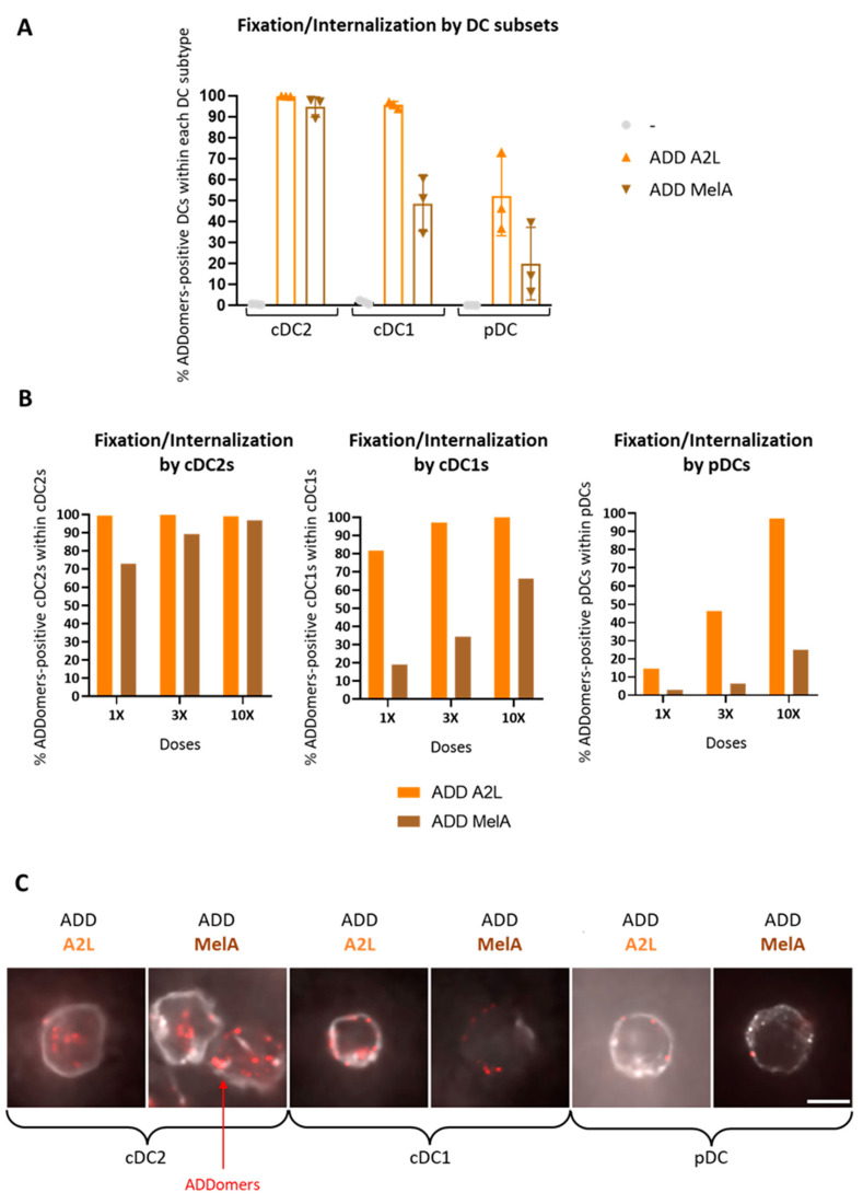
Virus-like particles constitute versatile vectors that can be used as vaccine platforms in many fields from infectiology and more recently to oncology. We previously designed non-infectious adenovirus-inspired 60-mer dodecahedric virus-like particles named ADDomers displaying on their surface either a short epitope or a large tumor/viral antigen. In this work, we explored for the first time the immunogenicity of ADDomers exhibiting melanoma-derived tumor antigen/epitope and their impact on the features of human dendritic cell (DC) subsets. We first demonstrated that ADDomers displaying tumor epitope/antigen elicit a strong immune-stimulating potential of human DC subsets (cDC2s, cDC1s, pDCs), which were able to internalize and cross-present tumor antigen, and subsequently cross-prime antigen-specific T-cell responses. To further limit off-target effects and enhance DC targeting, we engineered specific motifs to de-target epithelial cells and improve DCs' addressing. The improved engineered platform making it possible to display large antigen represents a tool to overcome the barrier of immune allele restriction, broadening the immune response, and paving the way to its potential utilization in humans as an off-the-shelf vaccine.

Keywords: C-type lectin receptors; adenovirus; immunotherapy; melanoma; vaccine platform.


Conflict of interest statement

The authors declare no conflict of interest.

Figures

Figure 1
Figure 1
Design and characterization of the different ADDomers. (A) Diagram showing the insertion of the A2L epitope (A2L in orange) and SpyTag (ST in pink) in the 2 loops of the ADDomer monomer. SpyTag can make an isopeptidic bond with SpyCatcher (SC in blue) fused to the RBD protein (in grey) or the MelanA protein (MelA in brown). (B) SDS-PAGE profiles of reduced and boiled samples of the different ADDomers showing the apparition of a higher MW covalent adduct for the ADD-MelA (ADD-ST/MelA-SC), ADD KGE RBD (ADD-KGE/RBD-SC) and ADD RGD RBD (ADD-RGD/RBD-SC). (C) Sequences of the MelA-SC and RBD-SC. In the MelA-SC sequence, the MelanA tumor antigen is colored in brown (A2L epitope is highlighted in orange), SpyCatcher is colored in blue and additional linkers and histidine tag are colored in black. In the RBD-SC sequence, the Spike Receptor Binding Domain of SARS-CoV-2 is colored in grey (N and O-glycosites are highlighted, respectively, in green and red), SpyCatcher is colored in blue and additional linkers and histidine tag are colored in black.
Figure 2
Figure 2
DC subsets efficiently fix and internalize ADDomers. PBMC (A,B) or PanDC (C) were incubated with different doses (1×, 3×, 10×) of fluorescent A2L/MelA-ADDomer for 1 h (A,B) or 2 h (C). The fixation and internalization of ADDomer by DC subsets were then evaluated by flow cytometry and confocal microscopy. (A) Percentage of fixation of ADDomer (3×) by each DC subset (n = 3 donors). (B) Dose response of ADDomer fixation by DC subsets (n = 1 donor). (C) Confocal imaging of ADDomer internalization by DC subsets. ADDomers are colored in red and the specific underlying BDCA1, BDCA2 or BDCA3 labeling is shown in gray. Scale bar is 5 microns and images are 18 × 18 microns.
Figure 3
Figure 3
ADDomer triggers a strong activation of DC subsets together with cytokine secretion. PBMCs were cultured for 24 h in control conditions (−), or with either a mixture of TLR-L (Poly IC, R848, CpGA), MelA26–35 peptide (10 μM), empty ADDomer (ADD Ø) or displaying A2L (ADD A2L) both at 1 μM. The viability, activation and cytokine secretion by DC subsets were then evaluated. (A) Impact of ADDomer on DCs’ viability assessed by measuring the percentage of alive cDC2s (BDCA1+), cDC1s (BDCA3+) and pDCs (BDCA2+) (n = 2 donors). (B) Impact of ADDomer on DCs’ activation depicted by evaluating the percentage of CD40+, CD80+ and CD86+ DC subsets (n = 2 donors). (C) Cytokine secretion measured in the culture supernatants by Luminex (n = 2 donors).
Figure 4
Figure 4
DCs efficiently cross-present the A2L epitope from ADDomer displaying MelA. Purified panDCs were incubated with ADD Ø, ADD A2L, ADD MelA, Peptide MelA and Peptide MelA + TLR-L for 1 h and cocultured 20 h with MelA26–35 specific CD8+ T cells. IFNg secretion was then evaluated by CBA. T cells were also cultured alone (−) or with PMA/iono (+) (n = 2 donors).
Figure 5
Figure 5
ADDomer displaying A2L epitope efficiently cross-prime peptide-specific CD8+ T cells dotted with a high affinity. PBMC were cultured in control conditions or with empty ADDomer (ADD Ø) or ADD A2L in the presence or not of a mixture of TLR-L. Cultures were restimulated every 7 days for 3 weeks. The elicitation of MelA-specific CD8 T cells was then evaluated by dextramer staining. (A) Evolution of Dext MelA+ CD8 T cells over time in the different conditions (n = 3 donors). (B) Amplification of Dext MelA+ CD8+ T cells over time. The amplification of antigen-specific CD8+ T cells was calculated based on the ratio of absolute numbers of dextramer+ CD8+ T cells at day X/day 0 (n = 3 donors). (C) Affinity of Dext MelA+ CD8+ T cells at day 20 (n = 3 donors).
Figure 6
Figure 6
ADD KGE and ADD RGD internalization by lung epithelial cell line A549. (A) ADD KGE AF 488 and ADD RGD AF 488 internalization by lung epithelial cell line A549 evaluated by flow cytometry at t = 20 min. (B) Time lapse imaging by confocal microscopy of ADD KGE AF 488 and ADD RGD AF 488 internalization by lung epithelial cell line A549. (C) Mean Fluorescence Intensity (MFI) of ADD KGE AF 488 and ADD RGD AF 488 within cells over time.
Figure 7
Figure 7
ADD KGE, ADD KGE RBD and RBD-SC binding evaluation on DC-SIGN and MGL receptors by surface plasmon resonance. (A) Sensorgrams of the interaction of the ADD-KGE constructs and RBD-SC onto DC-SIGN or MGL oriented surface. For ADD KGE and ADD KGE RBD, injected concentrations range from 136 nM to 0.2 nM by cascade dilution by a factor of 2. For RBD SC, concentration used range from 14.4 µM down to 56 nM by cascade dilution by 2. (B) Table of apparent KD calculated using steady state analysis in the BIAEval software.
Figure 8
Figure 8
ADD KGE/RGD (with or without RBD-SC and EDTA) fixation/internalization by DC subsets. PBMC pre-incubated or not with EDTA were incubated with fluorescent KGE/RBD ADDomer for 1 h. The fixation of ADDomer by DC subsets was then evaluated by flow cytometry. (A) Fixation and internalization evaluation of ADD KGE/RGD (with or without RBD-SC and EDTA) (0.5 µM i.e., 3×) on DC subsets using flow cytometry (n = 3 donors). (B) Fixation and internalization evaluation of ADD KGE/RGD (with or without RBD-SC and EDTA) (0.15 µM i.e., 1× or 0.5 µM i.e., 3× or 1.5 µM i.e., 10×) on DC subsets using flow cytometry (n = 1 donor).
Figure 9
Figure 9
DCs efficiently cross-present the A2L epitope only when displayed by ADD RGD. Purified panDCs were incubated with ADD Ø, ADD KGE/RGD +/− RBD, Peptide MelA and Peptide MelA + TLR-L for 1 h and cocultured 20 h with MelA26–35 specific CD8+ T cells. IFNg secretion was then evaluated by CBA. T cells were also cultured alone (−) or with PMA/iono (+) (n = 2 donors).

Similar articles

References

    1. Hu Z., Ott P.A., Wu C.J. Towards personalized, tumour-specific, therapeutic vaccines for cancer. Nat. Rev. Immunol. 2018;18:168–182. doi: 10.1038/nri.2017.131. - DOI - PMC - PubMed
    1. Galluzzi L., Vacchelli E., Bravo-San Pedro J.M., Buqué A., Senovilla L., Baracco E.E., Bloy N., Castoldi F., Abastado J.-P., Agostinis P., et al. Classification of current anticancer immunotherapies. Oncotarget. 2014;5:12472–12508. doi: 10.18632/oncotarget.2998. - DOI - PMC - PubMed
    1. Lichty B.D., Breitbach C.J., Stojdl D.F., Bell J.C. Going viral with cancer immunotherapy. Nat. Rev. Cancer. 2014;14:559–567. doi: 10.1038/nrc3770. - DOI - PubMed
    1. Chen D.S., Mellman I. Oncology meets immunology: The cancer-immunity cycle. Immunity. 2013;39:1–10. doi: 10.1016/j.immuni.2013.07.012. - DOI - PubMed
    1. Stevenson F.K., Palucka K. Understanding and activating immunity against human cancer. Curr. Opin. Immunol. 2010;22:212–214. doi: 10.1016/j.coi.2010.03.001. - DOI - PMC - PubMed

LinkOut - more resources