

Changing the Patient's Position: Pitfalls and Benefits for Radiation Dose and Image Quality of Computed Tomography in Polytrauma
- PMID: 36359504
- PMCID: PMC9689596
- DOI: 10.3390/diagnostics12112661
Changing the Patient's Position: Pitfalls and Benefits for Radiation Dose and Image Quality of Computed Tomography in Polytrauma
Abstract
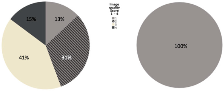
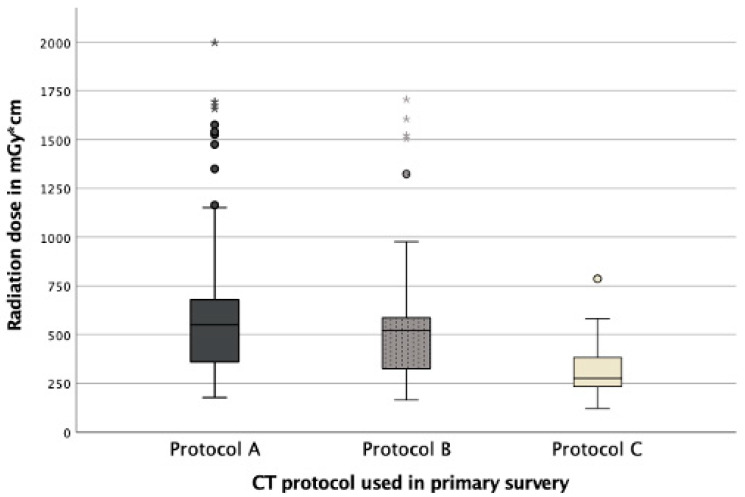
For computed tomography (CT), representing the diagnostic standard for trauma patients, image quality is essential. The positioning of the patient’s arms next to the abdomen causes artifacts and is also considered to increase radiation exposure. The aim of this study was to evaluate the effect of various positionings during different CT examination steps on the extent of artifacts as well as radiation dose using iterative reconstruction (IR). 354 trauma-CTs were analyzed retrospectively. All datasets were reconstructed using IR and three different examination protocols were applied. Arm elevation led to a significant improvement of the image quality across all examination protocols (p < 0.001). Variation in arm positioning during image acquisition did not lead to a reduction of radiation dose (p = 0.123). Only elevation during scout acquisition resulted in the reduction of radiation exposure (p < 0.001). To receive high-quality CT images, patients should be placed with elevated arms for the trunk scan, as artifacts remain even with the IR. Arm repositioning during the examination itself had no effect on the applied radiation dose because its modulation refers to the initial scout obtained. In order to achieve a dose effect by different positioning, a two-scout protocol (dual scout) should be used.
Keywords: computed tomography; image quality; iterative reconstruction; patient’s positioning; polytrauma; radiation exposure.
Conflict of interest statement
Jessica Graef, Keno Bressem and Patrick Asbach declare no conflict of interest. Stefan Niehues declares a relationship (honorary speaker) with the following company: Canon Medical Systems. Bernd Hamm declares relationships (institutional support) with the following companies: Abbott, Actelion Pharmaceuticals, Bayer Schering Pharma, Bayer Vital, BRACCO Group, Bristol Myers Squibb, Charité Research Organisation GmbH, Deutsche Krebshilfe, Dt. Stiftung für Herz- forschung, Essex Pharma, EU Programmes (Past President of ESR), Fibrex Medical Inc., Focused Ultrasound Surgery Foundation, Fraunhofer Gesellschaft, Guerbet, INC Research, InSightec Ltd., IPSEN Pharma, Kendle/MorphoSys AG, Lilly GmbH, Lundbeck GmbH, MeVis Medical Solutions AG, Nexus Oncology, Novartis, Parexel CRO Service, Perceptive, Pfizer GmbH, Phillips, sanofi-aventis S.A., Siemens, Spectranetics GmbH, Terumo Medical Corporation, TNS Healthcare GmbH, Toshiba, UCB Pharma, Wyeth Pharma, Zukunftsfonds Berlin (TSB).
Figures
Similar articles
-
Feasibility of radiation dose reduction with iterative reconstruction in abdominopelvic CT for patients with inappropriate arm positioning.PLoS One. 2018 Dec 31;13(12):e0209754. doi: 10.1371/journal.pone.0209754. eCollection 2018. PLoS One. 2018. PMID: 30596739 Free PMC article.
-
How does arm positioning of polytraumatized patients in the initial computed tomography (CT) affect image quality and diagnostic accuracy?Eur J Radiol. 2014 Jan;83(1):e67-71. doi: 10.1016/j.ejrad.2013.10.002. Epub 2013 Oct 16. Eur J Radiol. 2014. PMID: 24189387
-
Clinical evaluation of CT radiation dose in whole-body 18F-FDG PET/CT in relation to scout imaging direction and arm position.Ann Nucl Med. 2019 Mar;33(3):169-176. doi: 10.1007/s12149-018-1318-0. Epub 2018 Nov 10. Ann Nucl Med. 2019. PMID: 30415458
-
Basics of iterative reconstruction methods in computed tomography: A vendor-independent overview.Eur J Radiol. 2018 Dec;109:147-154. doi: 10.1016/j.ejrad.2018.10.025. Epub 2018 Oct 26. Eur J Radiol. 2018. PMID: 30527298 Review.
-
Intra-individual diagnostic image quality and organ-specific-radiation dose comparison between spiral cCT with iterative image reconstruction and z-axis automated tube current modulation and sequential cCT.Eur J Radiol Open. 2016 Jul 26;3:182-90. doi: 10.1016/j.ejro.2016.05.006. eCollection 2016. Eur J Radiol Open. 2016. PMID: 27504476 Free PMC article. Review.
References
-
- Piede D. Dosisreduktion: CT-Untersuchungen „fast ohne“ Dosis. Radiopraxis. 2015;8:19–28. doi: 10.1055/s-0034-1391572. - DOI
-
- Boas F.E., Fleischmann D. CT artifacts: Causes and reduction techniques. Imaging Med. 2012;4:229–240. doi: 10.2217/iim.12.13. - DOI
-
- Goeller A., Pogarell T., May M.S., Uder M., Dankerl P. Evaluation of CT-Guided Ultra-Low-Dose Protocol for Injection Guidance in Preparation of MR-Arthrography of the Shoulder and Hip Joints in Comparison to Conventional and Low-Dose Protocols. Diagnostics. 2021;11:1835. doi: 10.3390/diagnostics11101835. - DOI - PMC - PubMed
LinkOut - more resources
Full Text Sources
Research Materials
