

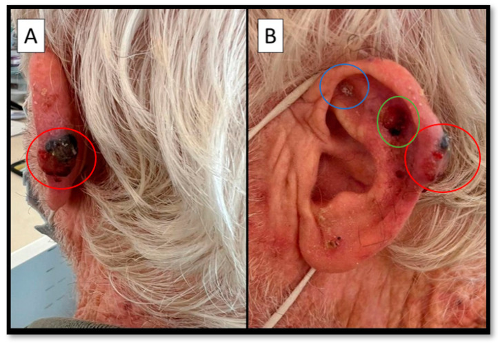
Polymorphic Malignant Melanoma (PMM) of the Left Helix: Case Report with Clinical-Pathological Correlations
- PMID: 36359557
- PMCID: PMC9688947
- DOI: 10.3390/diagnostics12112713
Polymorphic Malignant Melanoma (PMM) of the Left Helix: Case Report with Clinical-Pathological Correlations
Abstract
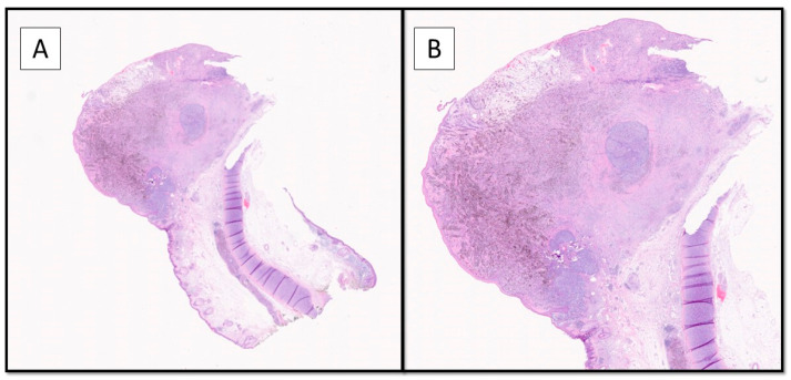
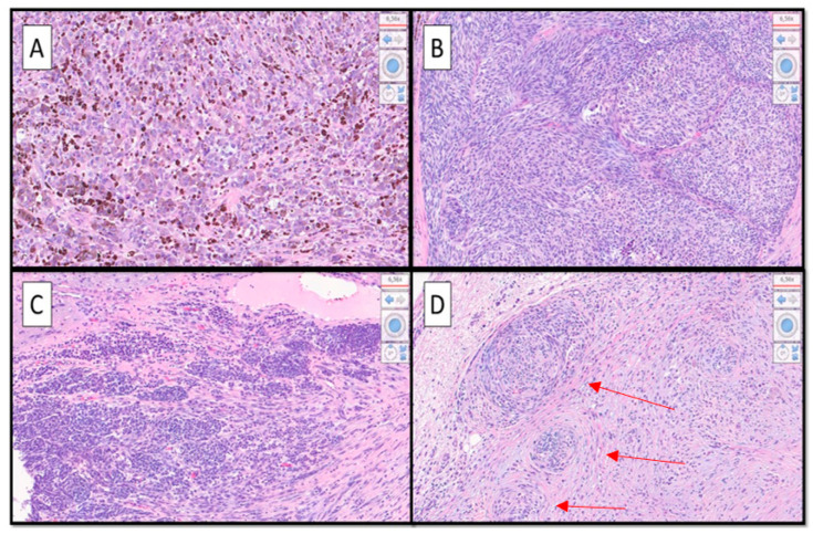
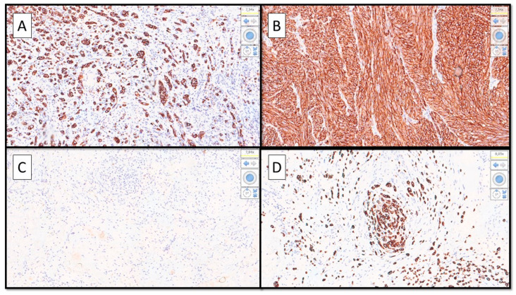
Malignant melanoma (MM) is known to be the great mimic in dermatopathology. Over time, several variants have been described, not all of which have repercussions on the clinical/oncological management of the affected patient. The existence, however, of these alternative forms of MM is of great interest to the pathologist, as they are potentially capable of inducing diagnostic errors affecting the diagnostic-therapeutic care pathway (PDTC). In this paper, we present a very rare case of polymorphic MM, in which five different morphological aspects coexisted in the same lesion, confirmed by immunohistochemical investigation and by RT-PCR for mutation of the BRAF gene and discuss the importance of correct recognition of these different morphological features to avoid misdiagnosis.
Keywords: differential diagnosis; immunohistochemistry; malignant melanoma; melanocytic; neoplasm; polymorphic; skin.
Conflict of interest statement
The authors declare no conflict of interest.
Figures
Similar articles
-
Role of In Vivo Reflectance Confocal Microscopy in the Analysis of Melanocytic Lesions.Acta Dermatovenerol Croat. 2018 Apr;26(1):64-67. Acta Dermatovenerol Croat. 2018. PMID: 29782304 Review.
-
Melanocytic Matricoma: A Potential Mimic of Malignant Melanoma.Cureus. 2023 Mar 30;15(3):e36907. doi: 10.7759/cureus.36907. eCollection 2023 Mar. Cureus. 2023. PMID: 37128540 Free PMC article.
-
CD44 and variants in melanocytic skin neoplasms.J Cutan Pathol. 1998 Apr;25(4):199-203. doi: 10.1111/j.1600-0560.1998.tb01719.x. J Cutan Pathol. 1998. PMID: 9609138
-
Partially Dedifferentiated Primitive Malignant Melanoma with Pseudo-Angiomatous Features: A Case Report with Review of the Literature.Diagnostics (Basel). 2023 Jan 29;13(3):495. doi: 10.3390/diagnostics13030495. Diagnostics (Basel). 2023. PMID: 36766604 Free PMC article.
-
Black and Brown Oro-facial Mucocutaneous Neoplasms.Head Neck Pathol. 2019 Mar;13(1):56-70. doi: 10.1007/s12105-019-01008-2. Epub 2019 Jan 29. Head Neck Pathol. 2019. PMID: 30693458 Free PMC article. Review.
References
-
- Cazzato G., Colagrande A., Cimmino A., Demarco A., Lospalluti L., Arezzo F., Resta L., Ingravallo G. The Great Mime: Three Cases of Melanoma with Carcinoid-Like and Paraganglioma-Like Pattern with Emphasis on Differential Diagnosis. Dermatopathology. 2021;8:130–134. doi: 10.3390/dermatopathology8020019. - DOI - PMC - PubMed
LinkOut - more resources
Full Text Sources
Research Materials
