. 2022 Oct 27;11(21):3405.
doi: 10.3390/cells11213405.

A Resilience Related Glial-Neurovascular Network Is Transcriptionally Activated after Chronic Social Defeat in Male Mice

Affiliations

A Resilience Related Glial-Neurovascular Network Is Transcriptionally Activated after Chronic Social Defeat in Male Mice

Constance Vennin et al. Cells. .

Abstract

Upon chronic stress, a fraction of individuals shows stress resilience, which can prevent long-term mental dysfunction. The underlying molecular mechanisms are complex and have not yet been fully understood. In this study, we performed a data-driven behavioural stratification together with single-cell transcriptomics of the hippocampus in a mouse model of chronic social defeat stress. Our work revealed that in a sub-group exhibiting molecular responses upon chronic stress, the dorsal hippocampus is particularly involved in neuroimmune responses, angiogenesis, myelination, and neurogenesis, thereby enabling brain restoration and homeostasis after chronic stress. Based on these molecular insights, we applied rapamycin after the stress as a proof-of-concept pharmacological intervention and were able to substantially increase stress resilience. Our findings serve as a data resource and can open new avenues for further understanding of molecular processes underlying stress response and for targeted interventions supporting resilience.

Keywords: cell-cell interaction; glial cells; hippocampus; neuroimmune pathways; neurovascular system; single-cell RNA-seq; stress resilience.


Conflict of interest statement

The authors declare no conflict of interest.

Figures
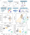
Figure 1
Figure 1
Behavioural classification of mice after chronic social defeat stress. (a) Overview of the experiments. Chronic social defeat stress was applied for 10 days with 3 times 15 s of fight per day. After 4 days, behavioural profiling was started. After the behavioural classification, three mice per stressed group (R, Int, S) and three non-stressed mice were selected. Brains were collected 2 days after the last behavioural test, and the dorsal and ventral hippocampus were dissected. Tissues were dissociated, and single and living cells were isolated by using a fluorescence-activated cell sorter. A total of 32,000 single-cells were captured by a droplet-based method and sequenced. Different cell populations were identified, the differentially expressed genes between the groups were analysed, and cell networks were uncovered. (b) Social interaction (SI) index of controls and defeated mice. After CSDS, two subgroups of defeated mice (highlighted in brown), i.e., the socially avoiding (SI < 0.75) and the socially non-avoiding (SI > 1.15) mice, were further subjected to a modified social interaction (MSI) test. (c) Based on their SI scores, the selected defeated mice highlighted in Figure 1b were clustered into three sub-phenotypes. Behavioural groups are visualized with principal component analysis, with four groups termed control (black), resilient (orange, R), intermediate (blue, Int), and susceptible (green, S) mice. The area around each cluster corresponds to the convex hull. (d) SI index of the four groups of mice. (e) SI index in the MSI test of the four groups of mice. Filled circles and triangles in (ce): individual mouse taken for subsequent single-cell RNA-seq experiment. * p < 0.05, ** p < 0.01 (Wilcoxon test in b, linear mixed-effects model followed by Tukey comparisons of marginal means in (e), # p < 0.05, ### p < 0.001 (Kruskal–Wallis test followed by Dunn’s posthoc comparisons in (d)).
Figure 2
Figure 2
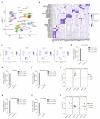
Single-cell analysis identifies different cell types in the different parts of the hippocampus as a specific response to chronic social defeat stress. (a) The uniform manifold approximation and projection (UMAP) plot of 29,358 cells in 41 clusters identified after thorough quality control. astros, astrocytes; OPC, oligodendrocytes progenitor cells; oligos, oligodendrocytes; micros, microglia; fibroblasts/micros, fibroblasts and microglia; micros/macros, microglia/macrophages; b-cells/micros, B lymphocytes/microglia. (b) Cell type annotation is based on the expression of well-known marker genes. The dot plot shows the expression of these genes across all the cell types. The dot size represents the percentage of cells expressing the gene, whereas the colour intensity of the dot corresponds to the average expression level. (c) The UMAP visualization of the 4 major cell populations, i.e., microglia, oligodendrocytes, endothelial and mural cells, showing the expression of representative well-known cell-type-specific marker genes. (dh) Number of differentially expressed genes of the resilient (R), intermediate (Int), and susceptible (S) mice compared to control mice and among all cell populations (d), microglia (e), oligodendrocytes (f), endothelial cells (g), mural cells (h) within the full hippocampus (hippocampus) and within the different parts: dorsal and ventral. (i,j) Differential expression of the immediate early genes (Arc, Egr1, c-Fos, Jun) (i) and the chronic social defeat stress marker genes (Hba-a1, Hba-a2, Hbb-bs) (j) of the resilient (R), the intermediate (Int) and the susceptible (S) mice compared to control mice in the entire hippocampus (hippocampus) and in the dissected parts of the hippocampus (dorsal, ventral). Each dot represents the average log2 fold change of differential expression of the corresponding gene between the two groups of mice in a given cell cluster.
Figure 3
Figure 3
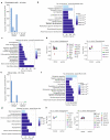
Chronic social defeat stress induces a microglia immune response in the hippocampal dorsal part of intermediate mice. (a) Number of DEGs among the microglial sub-clusters 1 to 8 in the intermediate mice compared to control mice in the dorsal and ventral parts of the hippocampus. (b) Top 10 GO terms analysis related to the DEGs identified in the dorsal hippocampus of the intermediate mice. The most relevant pathways are highlighted in bold. (c) Differential expression of M1 (Cxcl10, Fcgr2b, Fcgr3, H2-Aa, H2-D1, H2-Dmb1, H2-K1, H2-Oa, H2-Q4, H2-Q6, H2-T23, Tnfαip2, Tnfαip8l2, Il-1β) and M2 (Il-10rα, Socs3, Tgfβ1) markers in the dorsal and ventral parts of the hippocampus in the intermediate (Int) mice compared to control. (d) Difference in the percentage of cells (ΔPCT) expressing the M1 (Cxcl10, Fcgr2b, Fcgr3, H2-Aa, H2-D1, H2-Dmb1, H2-K1, H2-Oa, H2-Q4, H2-Q6, H2-T23, Tnfαip2, Tnfαip8l2, Il-1β) and M2 (Il-10rα, Socs3, Tgfβ1) markers in the dorsal and ventral parts of the hippocampus in the intermediate mice compared to control.
Figure 4
Figure 4
Chronic social defeat stress induces an oligodendrocyte response in the hippocampal dorsal part in the intermediate mice. (a) Number of DEGs among the oligodendrocytes sub-clusters 1 to 5 in the intermediate mice compared to control mice in the dorsal and ventral parts of the hippocampus. (b) Top 10 GO terms analysis related to the DEGs identified in the dorsal hippocampus of the intermediate mice. The most relevant pathways are highlighted in bold. (c) Differential expression of the oligodendrocyte progenitor (left panel), early and mature (right panel) oligodendrocyte markers in the intermediate (Int) mice compared to control mice in the dorsal and ventral parts of the hippocampus. (d) Differential expression of oligodendrocyte genes involved in microglia cell activation participating in the immune response pathway in the intermediate mice compared to control mice in the dorsal and ventral parts of the hippocampus. (e) Top 10 GO terms analysis related to the DEGs identified in the dorsal hippocampus of the sub-cluster micros_8 of the intermediate mice. The most interesting pathways are highlighted in bold. (f) Differential expression of the genes involved in the positive regulation of the glial cell differentiation pathway in the microglia cluster in the intermediate mice compared to control mice in the dorsal and ventral parts of the hippocampus. (g) Proposed cell-cell interaction identified in the intermediate mice between oligodendrocytes (top) and microglia (bottom).
Figure 5
Figure 5
Chronic social defeat stress induces an endothelial and mural cells response in the hippocampal dorsal part in the intermediate mice. (a) Number of DEGs among the endothelial sub-clusters 1 to 4 in the intermediate mice compared to control mice in the dorsal and ventral parts of the hippocampus. (b) Top 10 GO terms analysis related to the DEGs identified in the endothelial cells in the dorsal hippocampus of the intermediate mice. The most interesting pathways are highlighted in bold. (c) Selected GO terms related to the DEGs identified in the endothelial cells in the intermediate mice’s dorsal hippocampus. (d) Differential expression of the genes involved in the angiogenesis (left panel) and neurogenesis pathways (right panel) in the endothelial cells of intermediate (Int) mice compared to control mice in the dorsal and ventral parts of the hippocampus. (e) Number of DEGs among the mural sub-clusters 1 to 4 in the intermediate mice compared to control mice in the dorsal and ventral part of the hippocampus. (f) Top 10 GO terms analysis related to the DEGs identified in the mural cells in the dorsal hippocampus of the intermediate mice. The most interesting pathways are highlighted in bold. (g) Selected GO terms related to the DEGs identified in the mural cells in the dorsal hippocampus of the intermediate mice. (h) Differential expression of the genes involved in the angiogenesis and neurogenesis pathways in the mural cells of intermediate mice compared to control mice in the dorsal and ventral parts of the hippocampus.
Figure 6
Figure 6
A complex stress response to chronic social defeat in intermediate mice identifies a potential pharmacological target promoting stress resilience. (a) Circos plot representing the putative ligand receptor interactions between the brain cell types and upregulated in the dorsal hippocampus of intermediate mice. The edges are coloured according to the ligand-generating cell type. The thickness is proportional to the number of connections identified. (b) Ligand-receptor interaction network identified in the dorsal hippocampus of intermediate mice. The arrows point out from the ligands to the receptors. The colour of nodes corresponds to the brain cell types expressing the ligands and/or the receptors. (c) Differential expression of the matrix metalloproteinases (MMPs) inhibitors (upper panel) and extracellular matrix (ECM) components (lower panel) in the endothelial and mural cells of intermediate (Int) mice compared to control mice in the dorsal and ventral parts of the hippocampus. (d) Social interaction (SI) index of controls and defeated mice treated with vehicle or rapamycin after CSDS during 7 days. Rapamycin treatment after CSDS increases SI index of defeated animals and promotes stress resilience. (** p < 0.01 Two-way ANOVA followed by Tukey’s post hoc test.) (e) Distribution of the proportion of animals assigned to the three behavioural phenotypes (R, Int and S) in the different behavioural experiments performed in the present work: for the single-cell RNA-seq (scRNA), and for the pharmacological intervention (vehicle and rapamycin) experiment. A significant increase of resilient mice was observed in the rapamycin treated group compared to the non-treated group. (* p < 0.05 Chi squared test) (f) Overview of the proposed molecular mechanism observed at the blood-brain barrier. In defeated animals, the endogenous expression of MMPs inhibitors or the rapamycin treatment could promote stress resilience by maintaining the ECM integrity.

References

    1. Kalisch R., Baker D.G., Basten U., Boks M.P., Bonanno G.A., Brummelman E., Chmitorz A., Fernàndez G., Fiebach C.J., Galatzer-Levy I., et al. The Resilience Framework as a Strategy to Combat Stress-Related Disorders. Nat. Hum. Behav. 2017;1:784–790. doi: 10.1038/s41562-017-0200-8. - DOI - PubMed
    1. Kalisch R., Cramer A.O.J., Binder H., Fritz J., Leertouwer I.J., Lunansky G., Meyer B., Timmer J., Veer I.M., van Harmelen A.L. Deconstructing and Reconstructing Resilience: A Dynamic Network Approach. Perspect. Psychol. Sci. 2019;14:765–777. doi: 10.1177/1745691619855637. - DOI - PubMed
    1. Horn S.R., Feder A. Understanding Resilience and Preventing and Treating PTSD. Harv. Rev. Psychiatry. 2018;26:158–174. doi: 10.1097/HRP.0000000000000194. - DOI - PubMed
    1. Rakesh G., Morey R.A., Zannas A.S., Malik Z., Marx C.E., Clausen A.N., Kritzer M.D., Szabo S.T. Resilience as a Translational Endpoint in the Treatment of PTSD. Mol. Psychiatry. 2019;24:1268–1283. doi: 10.1038/s41380-019-0383-7. - DOI - PMC - PubMed
    1. Santomauro D.F., Mantilla Herrera A.M., Shadid J., Zheng P., Ashbaugh C., Pigott D.M., Abbafati C., Adolph C., Amlag J.O., Aravkin A.Y., et al. Global Prevalence and Burden of Depressive and Anxiety Disorders in 204 Countries and Territories in 2020 Due to the COVID-19 Pandemic. Lancet. 2021;398:1700–1712. doi: 10.1016/S0140-6736(21)02143-7. - DOI - PMC - PubMed

Publication types