Review
. 2022 Oct 28;11(21):3414.
doi: 10.3390/cells11213414.

Assaying Microglia Functions In Vitro

Affiliations
Review

Assaying Microglia Functions In Vitro

Emily Maguire et al. Cells. .

Abstract

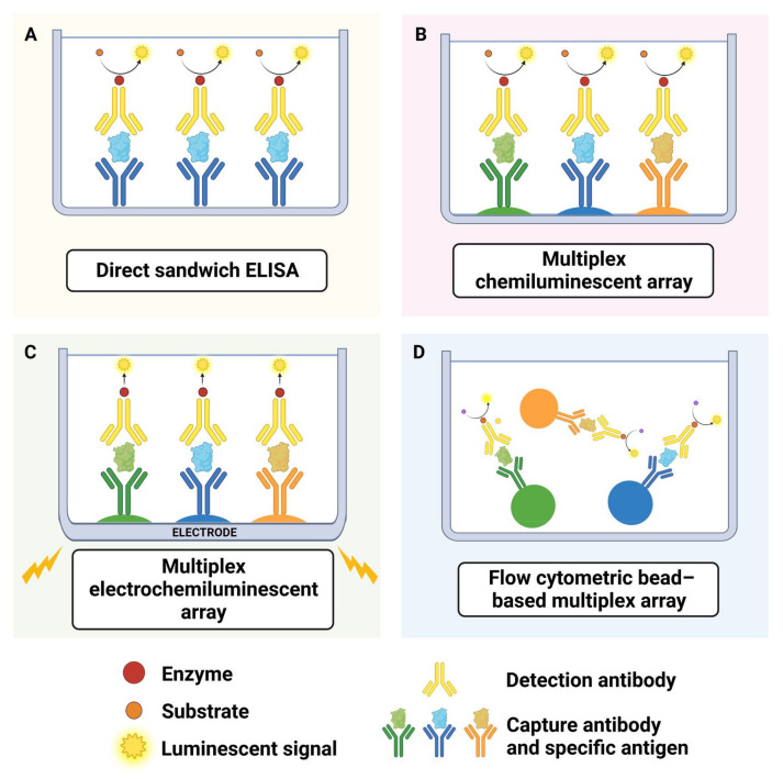
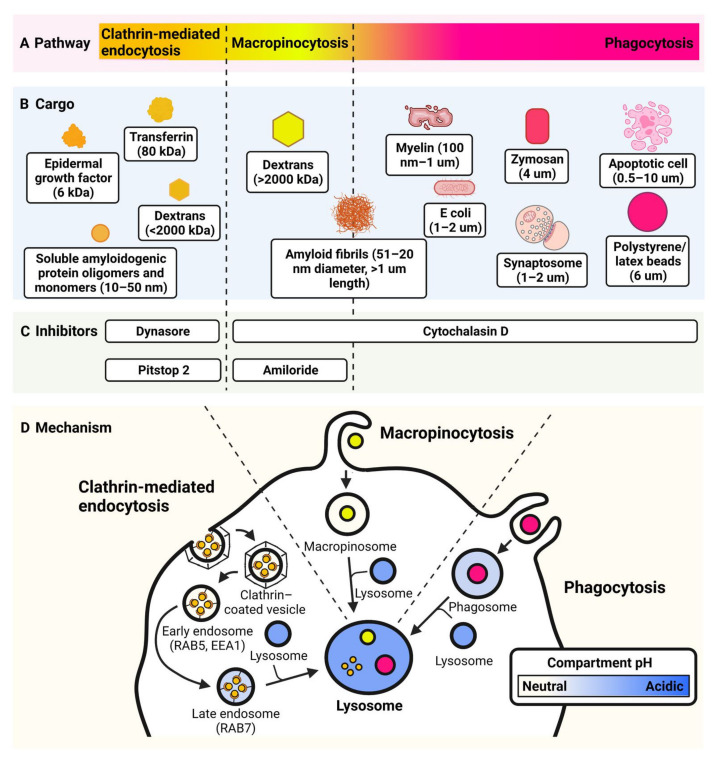
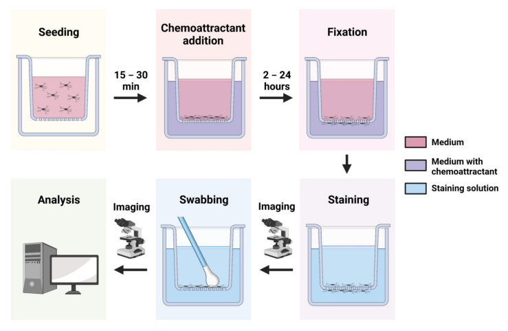
Microglia, the main immune modulators of the central nervous system, have key roles in both the developing and adult brain. These functions include shaping healthy neuronal networks, carrying out immune surveillance, mediating inflammatory responses, and disposing of unwanted material. A wide variety of pathological conditions present with microglia dysregulation, highlighting the importance of these cells in both normal brain function and disease. Studies into microglial function in the context of both health and disease thus have the potential to provide tremendous insight across a broad range of research areas. In vitro culture of microglia, using primary cells, cell lines, or induced pluripotent stem cell derived microglia, allows researchers to generate reproducible, robust, and quantifiable data regarding microglia function. A broad range of assays have been successfully developed and optimised for characterizing microglial morphology, mediation of inflammation, endocytosis, phagocytosis, chemotaxis and random motility, and mediation of immunometabolism. This review describes the main functions of microglia, compares existing protocols for measuring these functions in vitro, and highlights common pitfalls and future areas for development. We aim to provide a comprehensive methodological guide for researchers planning to characterise microglial functions within a range of contexts and in vitro models.

Keywords: chemotaxis; endocytosis; functional assays; iPSC; immunometabolism; in vitro; inflammation; microglia; motility; phagocytosis.


Conflict of interest statement

The authors declare no conflict of interest. The funders had no role in the design of the study; in the collection, analyses, or interpretation of data; in the writing of the manuscript; or in the decision to publish the results.

Figures

Figure 1
Figure 1
Overview of arrays for quantifying cytokine release. (A) ELISA: a 96 well plate is coated with a capture antibody specific to an antigen within the protein of interest, e.g., one specific cytokine/chemokine. Next, staining with another antibody that binds to the protein of interest and that is conjugated to an enzyme (e.g., streptavidin/biotin) allows detection/quantification of protein (Kohl and Ascoli, 2017). (B) Chemiluminescence arrays: multiple specific biotin-conjugated detection antibodies are added to each well, thus allowing quantification of multiple cytokines following the addition of horseradish peroxidase-conjugated streptavidin and measurement of luminescence. (C) Electrochemiluminescence arrays: each detection antibody, instead of being conjugated to biotin, is conjugated to a proprietary tag that is excited with emission beams in the electric field. (D) Bead based multiplex arrays: use proprietary bead sets that can be distinguished from each other via flow cytometry (due to varying size/fluorescence of bead types). Each type of bead comes conjugated to an antibody specific to one of the proteins of interest. Next, either streptavidin or fluorescence labelled antibodies are added that bind specifically to each cytokine-antibody complex on the bead sets. Using a flow cytometer, up to 25 cytokines in the same sample can be measured with commercial kits; or up to 100 with custom-conjugation [84].
Figure 2
Figure 2
Schematic of methods for assaying endocytosis and phagocytosis. (A,B) Pathways and cargoes of endocytosis. The size of particle determines the pathway utilised, indicated by colour on the schematic. Clathrin-mediated endocytosis (<120 nm) is shown in orange. Macropinocytosis (>200 nm) is shown in yellow. Phagocytosis (>500 nm) is shown in pink. (C) Inhibitors used in endocytic assays. (D) Mechanisms of endocytic pathways. Blue indicates relative acidity of compartments. The lysosome is pH 4.5–5.0. Endosomal protein biomarkers are also indicated.
Figure 3
Figure 3
Transwell assay workflow. Microglia are seeded into transwells and can be left to adhere to the membrane for up to 30 min. A defined chemoattractant is then added to the bottom chamber and microglia are allowed to migrate through the membrane for a defined timespan. Cells are fixed and stained. Light microscopy at a low magnification is used to image the inserts allowing visualization of all cells either side of the membrane. The inserts are then swabbed on the top side with a cotton wool bud to remove the unmigrated cells, and imaged again to visualise migrated cells only. Image analysis software is used to count cells, and the result is expressed as a ratio or percentage of migrated cells (after swabbing) relative to the total cell population (before swabbing).

Similar articles

Cited by

References

    1. Mittelbronn M., Dietz K., Schluesener H.J., Meyermann R. Local distribution of microglia in the normal adult human central nervous system differs by up to one order of magnitude. Acta Neuropathol. 2001;101:249–255. doi: 10.1007/s004010000284. - DOI - PubMed
    1. Tan Y.L., Yuan Y., Tian L. Microglial regional heterogeneity and its role in the brain. Mol. Psychiatry. 2020;25:351–367. doi: 10.1038/s41380-019-0609-8. - DOI - PMC - PubMed
    1. Menassa D.A., Muntslag T.A.O., Martin-Estebané M., Barry-Carroll L., Chapman M.A., Adorjan I., Tyler T., Turnbull B., Rose-Zerilli M.J.J., Nicoll J.A.R., et al. The spatiotemporal dynamics of microglia across the human lifespan. Dev. Cell. 2022;57:2127–2139.e6. doi: 10.1016/j.devcel.2022.07.015. - DOI - PMC - PubMed
    1. Nimmerjahn A., Kirchhoff F., Helmchen F. Resting microglial cells are highly dynamic surveillants of brain parenchyma in vivo. Science. 2005;308:1314–1318. doi: 10.1126/science.1110647. - DOI - PubMed
    1. Parakalan R., Jiang B., Nimmi B., Janani M., Jayapal M., Lu J., Tay S.S., Ling E.A., Dheen S.T. Transcriptome analysis of amoeboid and ramified microglia isolated from the corpus callosum of rat brain. BMC Neurosci. 2012;13:64. doi: 10.1186/1471-2202-13-64. - DOI - PMC - PubMed

Publication types

LinkOut - more resources