. 2022 Oct 28;11(21):3415.
doi: 10.3390/cells11213415.

Nogo-A Regulates the Fate of Human Dental Pulp Stem Cells toward Osteogenic, Adipogenic, and Neurogenic Differentiation

Affiliations

Nogo-A Regulates the Fate of Human Dental Pulp Stem Cells toward Osteogenic, Adipogenic, and Neurogenic Differentiation

Chai Foong Lai et al. Cells. .

Abstract

Human teeth are highly innervated organs that contain a variety of mesenchymal stem cell populations that could be used for cell-based regenerative therapies. Specific molecules are often used in these treatments to favorably modulate the function and fate of stem cells. Nogo-A, a key regulator of neuronal growth and differentiation, is already used in clinical tissue regeneration trials. While the functions of Nogo-A in neuronal tissues are extensively explored, its role in teeth still remains unknown. In this work, we first immunohistochemically analyzed the distribution of Nogo-A protein in the dental pulp of human teeth. Nogo-A is localized in a variety of cellular and structural components of the dental pulp, including odontoblasts, fibroblasts, neurons and vessels. We also cross-examined Nogo expression in the various pulp cell clusters in a single cell RNA sequencing dataset of human dental pulp, which showed high levels of expression in all cell clusters, including that of stem cells. We then assessed the role of Nogo-A on the fate of human dental pulp stem cells and their differentiation capacity in vitro. Using immunostaining, Alizarin Red S, Nile Red and Oil Red O staining we showed that Nogo-A delayed the differentiation of cultured dental pulp stem cells toward the osteogenic, adipogenic and neurogenic lineages, while addition of the blocking anti-Nogo-A antibody had opposite effects. These results were further confirmed by qRT-PCR, which demonstrated overexpression of genes involved in osteogenic (RUNX2, ALP, SP7/OSX), adipogenic (PPAR-γ2, LPL) and neurogenic (DCX, TUBB3, NEFL) differentiation in the presence of the anti-Nogo-A antibody. Conversely, the osteogenic and adipogenic genes were downregulated by Nogo-A. Taken together, our results show that the functions of Nogo-A are not restricted to neuronal cells but are extended to other cell populations, including dental pulp stem cells. We show that Nogo-A regulates their fates toward osteogenic, adipogenic and neurogenic differentiation, thus indicating its potential use in clinics.

Keywords: Nogo-A; adipogenic potential; anti-Nogo-A antibody; dental pulp; differentiation; human; human dental pulp stem cells; in vitro; neurogenic potential; osteogenic potential; single cell RNA sequencing; stem cell fates; tooth.


Conflict of interest statement

The authors declare no conflict of interest.

Figures

Figure 1
Figure 1
Experimental plan for analyzing the effects of Nogo-A (Nogo-A Δ20 fragment) and anti-Nogo-A antibody on the osteogenic, adipogenic, and neurogenic differentiation of human dental pulp stem cells.
Figure 2
Figure 2
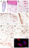
Nogo-A protein distribution in the dental pulp of human adult teeth and in cultured human dental pulp stem cells (hDPSCs). (A) Hematoxylin and Eosin (H&E) stained histological section of a human first premolar. (B,C) Histological sections of human adult teeth stained with H&E, showing the various components of the dentin-pulp complex. (DH) Immunostaining showing the distribution of Nogo-A (brown color) in odontoblasts (D,E), pulp fibroblasts (E,G,H), vessels (D,F,H) and nerves (E,F,G) of human teeth. Asterisks in (H) indicate endothelial cells. (I) Nogo-A immunostaining (brown/red color) in cultured hDPSCs. (J) Nogo-A immunofluorescence staining (red color) in hDPSCs. DAPI (blue color) marks the nuclei of the cells. Abbreviations: d, dentin; e, enamel; n, nerves; o, odontoblasts; p, dental pulp; v, vessels.
Figure 3
Figure 3
Feature plots showing the expression of NOGO and NOGO-related receptors genes in the dental pulp stem cell compartment of human adult teeth. (A) Specific cell clusters within the dental pulp. In blue the DPSCs cluster. (BG) Cells expressing RTN4 (NOGO; (B)), LINGO1 (C), RTN4R (D), S1PR2 (E), TNFRF19 (F), and NGFR (G) in all dental pulp compartments. Expression in individual cells is represented by red dots. (H) Quantitative details of the NOGO and NOGO-related receptors genes within DPSCs cluster.
Figure 4
Figure 4
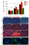
Effects of the Nogo-A protein (Nogo-A Δ20 fragment) and anti-Nogo-A antibody on the osteogenic differentiation of hDPSCs. (A) Quantification of Alizarin Red S staining in hDPSCs cultured in osteogenic medium (Os) alone (black colored columns), in osteogenic medium supplemented with the anti-Nogo-A antibody (red colored columns), and in osteogenic medium supplemented with the Nogo-A Δ20 protein (green colored columns) for 7, 14 and 21 days. Four independent biological replicates were used. The bars represent the means ± standard errors. Asterisks above the error bars denote significant differences (*, p < 0.05; **, p < 0.01; ***, p < 0.001) by one-way analysis of variance (ANOVA). (BJ) Alizarin Red S staining indicating the formation of mineralized nodules by hDPSCs cultured in the osteogenic medium alone (B,E,H), in osteogenic medium supplemented with the anti-Nogo-A antibody (C,F,I), and in osteogenic medium supplemented with the Nogo-A Δ20 protein (D,G,J) for 7, 14 and 21 days. (KM) Collagen type-I immunofluorescence staining (green color) in hDPSCs cultured for 21 days in the osteogenic medium alone (K), in osteogenic medium supplemented with the anti-Nogo-A antibody (L), and in osteogenic medium supplemented with the Nogo-A Δ20 protein (M). DAPI (blue color) marks the cell nuclei. Abbreviation: n, mineralization nodules. Scale bars are as indicated.
Figure 5
Figure 5
Effects the Nogo-A protein and anti-Nogo-A antibody on the expression of genes during hDPSCs osteogenic differentiation. qRT-PCR analysis showing THY1/CD90 (A), END/CD105 (B), RUNX2 (C), ALP (D), and SP7/OSX (E) expression levels in hDPSCs cultured in osteogenic medium (Os) alone (black columns), in osteogenic medium supplemented with the anti-Nogo-A-antibody (red colored columns), and osteogenic medium supplemented with the Nogo-A Δ20 protein (green colored columns) for 7, 14 and 21 days. Values, normalized to Glyceraldaeyde-3-Phosphate Dehydrogenase (GAPDH), shown are the mean ± SD of triplicates. Statistical analysis used Student’s t-test (significant difference at *, p < 0.05; **, p < 0.01).
Figure 6
Figure 6
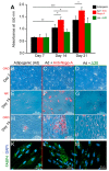
Effects of the Nogo-A protein (Nogo-A Δ20 fragment) and anti-Nogo-A antibody on the adipogenic differentiation of hDPSCs. (A) Quantification of Oil Red O (ORO) staining in hDPSCs cultured in adipogenic medium (Ad) alone (black colored columns), adipogenic medium supplemented with the anti-Nogo-A antibody (red colored columns), and adipogenic medium supplemented with the Nogo-A Δ20 protein (green colored columns) for 7, 14 and 21 days. Four independent biological replicates were used. The bars represent the means ± standard errors. Asterisks above the error bars denote significant differences (*, p < 0.05; **, p < 0.01; ***, p < 0.001) by one-way analysis of variance (ANOVA). (BJ) ORO (BD,HJ) and Nile Red (NR) staining (EG) indicating the formation of lipid droplets (red color) in hDPSCs cultured in adipogenic medium alone (B,E,H), in adipogenic medium supplemented with the anti-Nogo-A antibody (C,F,I), and in adipogenic medium supplemented with the Nogo-A Δ20 protein (D,G,J) for 7 and 14 days. (KM) Fatty Acid Binding Protein 4 (FABP4) immunofluorescence staining (green color) in hDPSCs cultured for 21 days in adipogenic medium alone (K), in adipogenic medium supplemented with the anti-Nogo-A antibody (L), and in adipogenic medium supplemented with the Nogo-A Δ20 protein (M). DAPI (blue color) marks the nuclei of the cells. Scale bars are as indicated.
Figure 7
Figure 7
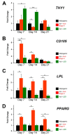
Effects of the Nogo-A protein and anti-Nogo-A antibody on the expression of genes during hDPSC adipogenic differentiation. qRT-PCR analysis of the relative expression levels of THY1/CD90 (A), END/CD105 (B), LPL (C) and PPARG (D) in hDPSCs cultured in adipogenic medium (Ad) alone (black columns), in adipogenic medium supplemented with the anti-Nogo-A-antibody (red colored columns), and in adipogenic medium supplemented with the Nogo-A Δ20 protein (green colored columns) for 7, 14 and 21 days. Values, normalized to GAPDH, shown are the mean ± SD of triplicates. Statistical analysis used Student’s t-test (significant difference at *, p < 0.05; **, p < 0.01).
Figure 8
Figure 8
Effects of the Nogo-A protein and anti-Nogo-A antibody on hDPSCs neuronal differentiation. (AC) Morphological changes of hDPSCs cultured in neurogenic induction medium (Neur) alone (A), in neurogenic medium supplemented with the anti-Nogo-A-antibody (B), and in neurogenic medium supplemented with the Nogo-A Δ20 protein (C) for 14 days. (DF) Neurofilament-L (NFL) immunofluorescent staining (green color) in hDPSCs cultured for 7 days in neurogenic medium alone (D), in neurogenic medium supplemented with the anti-Nogo-A-antibody (E), and in neurogenic medium supplemented with the Nogo-A Δ20 protein (F). DAPI (blue color) marks cell nuclei. (GI) Tubulin Beta-III (TUBB3) immunofluorescent staining (green color) in hDPSCs cultured for 7 days in neurogenic medium alone (G), in neurogenic medium supplemented with the anti-Nogo-A-antibody (H), and in neurogenic medium supplemented with the Nogo-A Δ20 protein (I). DAPI in blue color.
Figure 9
Figure 9
Effects the Nogo-A protein and anti-Nogo-A antibody on the expression of genes during hDPSC neuronal differentiation. qRT-PCR analysis of the relative expression levels of THY1/CD90 (A), END/CD105 (B), TUBB3 (Tubulin Beta-III; (C)), DCX (Doublecortin; (D)) and NFL (Neurofilament-L; (E)) in hDPSCs cultured in neurogenic medium (Neur) alone (black columns), in neurogenic medium supplemented with the anti-Nogo-A antibody (red color columns), and in neurogenic medium supplemented with the Nogo-A Δ20 protein (green color columns) for 7, 14 and 21 days. Values, normalized to Glyceraldaeyde-3-Phosphate Dehydrogenase (GAPDH), shown are the mean ± SD of triplicates. Statistical analysis used Student’s t-test (significant difference at *, p < 0.05; **, p < 0.01).
Figure 10
Figure 10
Illustration depicting the effects of Nogo-A and anti-Nogo-A-antibody on the hDPSC differentiation into adipogenic, osteogenic and neurogenic lineages. Based on our hypothesis, in osteogenic and neurogenic microenvironments hDPSCs derived from the THY1-positive cell lineage (yellow color) enhance their differentiation potential toward either osteoblasts or neurons in absence of Nogo-A. In adipogenic microenvironment, hDPSCs originated from the CD105-positive cells (light green color) are directed toward an adipocytic fate in the absence of Nogo-A. Dental pulp fibroblasts in dark green color, nerves in blue color, vessels in red color. The territory depicted with the blue/green color indicates areas with various levels of Nogo-A protein expression.

Similar articles

Cited by

References

    1. Pera M.F., Tam P.P. Extrinsic regulation of pluripotent stem cells. Nature. 2010;465:713–720. doi: 10.1038/nature09228. - DOI - PubMed
    1. Nombela-Arrieta C., Ritz J., Silberstein L.E. The elusive nature and function of mesenchymal stem cells. Nat. Rev. Mol. Cell Biol. 2011;12:126–131. doi: 10.1038/nrm3049. - DOI - PMC - PubMed
    1. Signer R.A., Morrison S.J. Mechanisms that regulate stem cell aging and life span. Cell Stem Cell. 2013;12:152–165. doi: 10.1016/j.stem.2013.01.001. - DOI - PMC - PubMed
    1. Wells J.M., Watt F.M. Diverse mechanisms for endogenous regeneration and repair in mammalian organs. Nature. 2018;557:322–328. doi: 10.1038/s41586-018-0073-7. - DOI - PubMed
    1. Mitsiadis T.A., Barrandon O., Rochat A., Barrandon Y., De Bari C. Stem cell niches in mammals. Exp. Cell Res. 2007;313:3377–3385. doi: 10.1016/j.yexcr.2007.07.027. - DOI - PubMed

Publication types