Review
. 2022 Oct 29;11(21):3421.
doi: 10.3390/cells11213421.

The Hidden Role of Non-Canonical Amyloid β Isoforms in Alzheimer's Disease

Affiliations
Review

The Hidden Role of Non-Canonical Amyloid β Isoforms in Alzheimer's Disease

Lukas Busch et al. Cells. .

Abstract

Recent advances have placed the pro-inflammatory activity of amyloid β (Aβ) on microglia cells as the focus of research on Alzheimer's Disease (AD). Researchers are confronted with an astonishing spectrum of over 100 different Aβ variants with variable length and chemical modifications. With the exception of Aβ1-42 and Aβ1-40, the biological significance of most peptides for AD is as yet insufficiently understood. We therefore aim to provide a comprehensive overview of the contributions of these neglected Aβ variants to microglia activation. First, the impact of Aβ receptors, signaling cascades, scavenger mechanisms, and genetic variations on the physiological responses towards various Aβ species is described. Furthermore, we discuss the importance of different types of amyloid precursor protein processing for the generation of these Aβ variants in microglia, astrocytes, oligodendrocytes, and neurons, and highlight how alterations in secondary structures and oligomerization affect Aβ neurotoxicity. In sum, the data indicate that gene polymorphisms in Aβ-driven signaling pathways in combination with the production and activity of different Aβ variants might be crucial factors for the initiation and progression of different forms of AD. A deeper assessment of their interplay with glial cells may pave the way towards novel therapeutic strategies for individualized medicine.

Keywords: APP; Alzheimer’s Disease; Amyloid Beta; cell-surface receptors; glia; microglia; neurodegeneration; neuroinflammation.


Conflict of interest statement

The authors declare no conflict of interest.

Figures
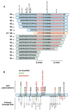
Figure 2
Figure 2
Truncated and modified Aβ species. (A): Depiction of N- and C-truncated Aβ variants and their potential modification sites as described in Kummer and Heneka, 2014. Motifs for β-sheet formation are indicated in red (race = racemization, iso = isomerization, pyro = pyroglutamylation, phos = phosphorylation, glycol = glycosylation, nitra = nitration, ox = oxidation). (B): Aβ peptide with polymorphisms of APP that directly influence the amino acid composition of its Aβ domain (up) [86] and the proposed cleavage sites of various secretases and enzymes (down) [11]. Polymorphisms are classified according to their proposed pathological influence [86]. NEP = neprilysin, PLG = plasmin, ACE = angiotensin converting Enzyme, IDE = insulin-degrading enzyme, M2 = MMP-2, M9 = MMP-9, ECE1/2 = endothelin converting enzyme 1/2, β2 = BACE2, CATB = cathepsin B.
Figure 1
Figure 1
Membrane topology and domain architecture of microglial Aβ-binding receptors. Some receptors are shown exemplarily to underpin the diverging domain architecture und potential Aβ-binding site positions as far as identified (red arrows). Relative location of polymorphisms to binding sites are indicated by yellow diamonds. Proportion of domains has been neglected. For RAGE, binding of Aβ is assumed to occur as a dimer on the dimeric receptor [16]. In addition to cleaved soluble RAGE and TREM2, additional soluble splice forms exist [17,18].
Figure 3
Figure 3
The amyloidogenic and non-amyloidogenic pathway of APP processing. (A) The non-amyloidogenic pathway is starting by α-secretase cleavage of APP which leads to the release of soluble APPα (sAPPα) and concomitant generation of the α-C-terminal fragment (α-CTF). Subsequent cleavage of the α-CTF by γ-secretase leads to the secretion of the small fragment p3 and release of the APP intracellular C-terminal domain (AICD) into the cytosol. In the amyloidogenic pathway, APP is first cleaved by β-secretase to produce soluble APPβ (sAPPβ) and the membrane retained β-C-terminal fragment (β-CTF). Further processing of the β-CTF by γ-secretase releases AICD as well as the Aβ peptide, which can form neurotoxic oligomers. Aβ production was reported to be higher in neuronal cultures compared to astrocytes or microglia cultures [95,96]. 3D structures (PDB: 1IYT) are based on NMR experiments by Crescenzi and colleagues [97]. (B) Glial cells mainly produce N-abridged Aβ peptides (up to 60%) such as Aβ2/3-X or Aβ4/5-x, while neurons produce predominantly Aβ peptides starting at position 1 (80%) [98].
Figure 4
Figure 4
Cellular responses of microglia after detection of Aβ. Microglia are capable of perceiving soluble Aβ peptides and insoluble aggregates via multiple cell-surface receptors, which then induce varied cellular responses such induction of cell migration, cytokine and chemokine release, secretion of proteases, and generation of oxidative stress [3,23]. These processes are in part regulated by the intracellular NLRP3 inflammasome. The release of inflammasome components (ASC specks) may lead to seeding of Aβ and may thus contribute to plaque formation [248].
Figure 5
Figure 5
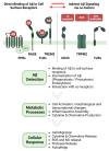
Aβ-mediated activation of microglia. Direct detection of Aβ (e.g., by TREM2, TLRs, FPRs, RAGE) or indirect stimulation through Aβ-mediated processes (e.g., TRPM2 activation by ROS, detection of autoantibodies against Aβ by FcRs) leads microglia to adopt their reactive phenotype in which they utilize intracellular signaling pathways and metabolic processes to initiate their pro-inflammatory response.

References

    1. Akiyama H., Arai T., Kondo H., Tanno E., Haga C., Ikeda K. Cell mediators of inflammation in the Alzheimer disease brain. Alzheimer Dis. Assoc. Disord. 2000;14((Suppl. 1)):S47–S53. doi: 10.1097/00002093-200000001-00008. - DOI - PubMed
    1. Heneka M.T., Kummer M.P., Latz E. Innate immune activation in neurodegenerative disease. Nat. Rev. Immunol. 2014;14:463–477. doi: 10.1038/nri3705. - DOI - PubMed
    1. Leng F., Edison P. Neuroinflammation and microglial activation in Alzheimer disease: Where do we go from here? Nat. Rev. Neurol. 2021;17:157–172. doi: 10.1038/s41582-020-00435-y. - DOI - PubMed
    1. Rogers J., Luber-Narod J., Styren S.D., Civin W.H. Expression of immune system-associated antigens by cells of the human central nervous system: Relationship to the pathology of Alzheimer’s disease. Neurobiol. Aging. 1988;9:339–349. doi: 10.1016/S0197-4580(88)80079-4. - DOI - PubMed
    1. McGeer P.L., Itagaki S., Tago H., McGeer E.G. Occurrence of HLA-DR reactive microglia in Alzheimer’s disease. Ann. N. Y. Acad. Sci. 1988;540:319–323. doi: 10.1111/j.1749-6632.1988.tb27086.x. - DOI - PubMed

Publication types

MeSH terms