Review
. 2022 Oct 30;11(21):3429.
doi: 10.3390/cells11213429.

Bioengineering Human Pluripotent Stem Cell-Derived Retinal Organoids and Optic Vesicle-Containing Brain Organoids for Ocular Diseases

Affiliations
Review

Bioengineering Human Pluripotent Stem Cell-Derived Retinal Organoids and Optic Vesicle-Containing Brain Organoids for Ocular Diseases

Peggy Arthur et al. Cells. .

Abstract

Retinal organoids are three-dimensional (3D) structures derived from human pluripotent stem cells (hPSCs) that mimic the retina's spatial and temporal differentiation, making them useful as in vitro retinal development models. Retinal organoids can be assembled with brain organoids, the 3D self-assembled aggregates derived from hPSCs containing different cell types and cytoarchitectures that resemble the human embryonic brain. Recent studies have shown the development of optic cups in brain organoids. The cellular components of a developing optic vesicle-containing organoids include primitive corneal epithelial and lens-like cells, retinal pigment epithelia, retinal progenitor cells, axon-like projections, and electrically active neuronal networks. The importance of retinal organoids in ocular diseases such as age-related macular degeneration, Stargardt disease, retinitis pigmentosa, and diabetic retinopathy are described in this review. This review highlights current developments in retinal organoid techniques, and their applications in ocular conditions such as disease modeling, gene therapy, drug screening and development. In addition, recent advancements in utilizing extracellular vesicles secreted by retinal organoids for ocular disease treatments are summarized.

Keywords: assembled organoids; extracellular vesicles; human induced pluripotent stem cells; ocular diseases; retinal organoids.


Conflict of interest statement

The authors declare no conflict of interest.

Figures

Figure 1
Figure 1
Schematic summary of applications of brain and retinal organoids derived from human pluripotent stem cells.
Figure 2
Figure 2
Summary of various protocols (AE) for retinal organoid differentiation from human induced pluripotent stem cells. (A). The protocol of using 3D–2D–3D culture in retinal differentiation medium. Chichagova et al., 2017 [43]. (B). The protocol of using 2D–3D culture in ProB27 medium. Reichman et al., 2019 [44], (C) Long-term retinal differentiation with embryoid bodies, which shows the photoreceptor maturation. Kaya et al., 2019 [45]. (D) The protocol of using 2D–3D culture in NSC medium. Zhu et al., 2018 [46]. (E) Long-term retinal differentiation starting with cyst and adherent culture for organoid maturation. Kim et al., 2019 [47]. Neural induction medium (NIM), retinal differentiation medium (RDM), retinal maintenance medium (RMM), fetal bovine serum (FBS), photoreceptor induction medium (PIM), fibroblast growth factor (FGF), neural stem cell (NSC).
Figure 3
Figure 3
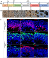
Retinal organoid differentiation and characterization [68]. (A) Schematic illustration showing the differentiation of retinal organoids from hiPSC and timeline representation of the early (Day 30–120) and late organoids (Day 121–300) for exosome preparation. (B) Phase contrast microscopic images showing the morphology of retinal organoids at different time points of differentiation (Day 2, B1; Day 25, B2; Day 45, B3; Day 200, B4). Scale bar, 200 µm. (C) Confocal images of 90-day old retinal organoids (C1–C8) stained for retinal progenitor cells (Pax6, C1; Vx2, C2), ganglion and amacrine cells (Brn3a, C3), pan photoreceptors (Crx, C4; Rcvrn, C5; Aipl1; C6), rod photoreceptor (NRL; C7), and cone photoreceptor (ARR3, C8). Image of 200-day old retinal organoids showing the staining of rhodopsin protein indicating the maturation of rod photoreceptor (Rho, C9). Scale bar, 50 µm. Figure reproduced from Arthur et al., (2022) under a Creative Commons license.
Figure 4
Figure 4
Differentiation protocols of optic vesicle-containing brain organoids for ocular applications. (A) Differentiation of iPSCs to brain organoids with optic vesicle regions (OVB), Elke et al., 2021 [83]. (B) Differentiation of iPSCs to complex organoid models created by the generation of both retinal and forebrain cortical organoids, Milan et al., 2022 [23]. These organoids can help to study brain–eye interactions during embryo development, model congenital retinal disorders, and generate patient-specific retinal cell types for personalized drug testing and transplantation therapies.
Figure 5
Figure 5
Schematic illustration of various applications of brain and retinal organoids and their derived exosomes in ocular diseases.

Similar articles

Cited by

References

    1. Huch M., Koo B.-K. Modeling mouse and human development using organoid cultures. Development. 2015;142:3113–3125. doi: 10.1242/dev.118570. - DOI - PubMed
    1. Lancaster M.A., Knoblich J.A. Organogenesis in a dish: Modeling development and disease using organoid technologies. Science. 2014;345:1247125. doi: 10.1126/science.1247125. - DOI - PubMed
    1. Corrò C., Novellasdemunt L., Li V.S. A brief history of organoids. Am. J. Physiol. Cell Physiol. 2020;319:C151–C165. doi: 10.1152/ajpcell.00120.2020. - DOI - PMC - PubMed
    1. Ootani A., Li X., Sangiorgi E., Ho Q.T., Ueno H., Toda S., Sugihara H., Fujimoto K., Weissman I.L., Capecchi M.R., et al. Sustained in vitro intestinal epithelial culture within a Wnt-dependent stem cell niche. Nat. Med. 2009;15:701–706. doi: 10.1038/nm.1951. - DOI - PMC - PubMed
    1. Xu H., Jiao Y., Qin S., Zhao W., Chu Q., Wu K. Organoid technology in disease modelling, drug development, personalized treatment and regeneration medicine. Exp. Hematol. Oncol. 2018;7:30. doi: 10.1186/s40164-018-0122-9. - DOI - PMC - PubMed

Publication types

LinkOut - more resources