Review
. 2022 Oct 31;11(21):3439.
doi: 10.3390/cells11213439.

The DNA Methylation in Neurological Diseases

Affiliations
Review

The DNA Methylation in Neurological Diseases

Samareh Younesian et al. Cells. .

Abstract

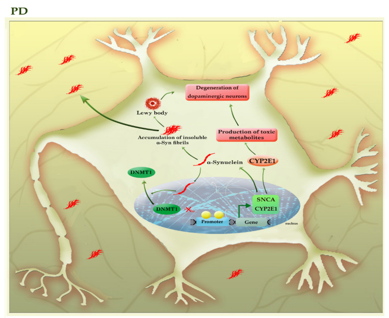
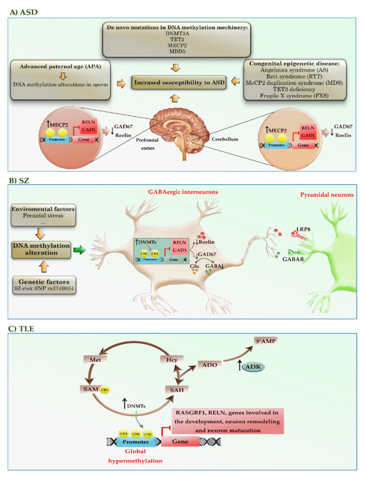
DNA methylation is critical for the normal development and functioning of the human brain, such as the proliferation and differentiation of neural stem cells, synaptic plasticity, neuronal reparation, learning, and memory. Despite the physical stability of DNA and methylated DNA compared to other epigenetic modifications, some DNA methylation-based biomarkers have translated into clinical practice. Increasing reports indicate a strong association between DNA methylation profiles and various clinical outcomes in neurological diseases, making DNA methylation profiles valuable as novel clinical markers. In this review, we aim to discuss the latest evidence concerning DNA methylation alterations in the development of neurodegenerative, neurodevelopmental, and neuropsychiatric diseases. We also highlighted the relationship of DNA methylation alterations with the disease progression and outcome in many neurological diseases such as Alzheimer's disease, Parkinson's disease, amyotrophic lateral sclerosis, frontotemporal dementia, and autism.

Keywords: Alzheimer’s disease; DNA methylation; Huntington’s disease; Parkinson’s disease; autism; neurological disorders.


Conflict of interest statement

The authors declare no conflict of interest.

Figures

Figure 1
Figure 1
A schematic representation of the selected mechanisms in the pathogenesis of FXS. The presence of more than 200 copies of CGG repeat in the 5′ UTR region of the FMR1 gene leads to promoter hypermethylation, transcriptional silencing, and loss of FMR protein. mGluRI: group 1 metabotropic glutamate receptors; GABA: gamma-aminobutyric acid.
Figure 2
Figure 2
A schematic representation of the selected mechanisms in the pathogenesis of HD. HTT gene, located at 4p16, has 64 exons and a CAG trinucleotide repeat expansion in the exon 1 region. The CAG repeats in the coding sequence of mutant HTT (mHTT) produce mHTT exon 1 protein. mHTT exon 1 protein enters the nucleus. The gradual aggregation of mHTT exon 1 oligomers leads to the formation of large inclusions in the cytoplasm and nucleus of neural cells. The intracytoplasmic inclusions (ICIs) have various toxic effects on neural cells and can exacerbate the aggregation of mHTT exon 1 protein. Whereas the polyQ tract of intranuclear inclusions (INIs) recruits MECP2 to the promoter of BDNF, downregulating the expression of BDNF. BDNF: brain-derived neurotrophic factor; Cyt-c: cytochrome c; mHTT: mutant huntingtin; NMDAR: N-methyl-D-aspartate receptor; ROS: reactive oxygen species.
Figure 3
Figure 3
A schematic representation of the selected mechanisms in the pathogenesis of C9-ALS/FTD. Loss of function; the presence of pathogenic hexanucleotide G4C2 repeats expansion (pHRE) within the promoter region inhibits transcription processes and reduces C9orf72 protein levels. Gain of function; the presence of pHRE within intron 1 leads to the sequestration of RNA binding proteins (RBPs) and the accumulation of toxic RNA foci and dipeptide repeat proteins (DRPs). The promoter hypermethylation of the C9orf72 gene reduces the accumulation of RNA foci and/or DRPs aggregation in the neural cells. Amino acid abbreviations: A: alanine, R: arginine, G: glycine, and P: proline.
Figure 4
Figure 4
A schematic representation of the selected mechanisms in the pathogenesis of AD. Under physiological conditions, APP is cleaved in the non-amyloidogenic pathway (not shown). In the absence of SORL1 due to epigenetic silencing or mutation, APP is shunted into the late endosomal pathway. In the amyloidogenic pathway, APP enters the late endosome, where it is cleaved by the β-secretase (BACE1), and then by γ-secretase. AICD enters the nucleus and acts as a transcription factor, whereas the Aβ40/42 peptides and sAPPβ are secreted to the extracellular space. An imbalance of Aβ production and its clearance from the brain promotes Aβ aggregation and deposition. The Aβ aggregates can activate the kinases involved in the tau pathway, leading to tau hyperphosphorylation. The aberrant hyperphosphorylation of tau causes p-tau to be separated from microtubules (MTs), leading to MTs depolymerization and axonal degeneration. The disruption of the tau pathway leads to the accumulation of tau aggregates to form oligomers and neurofibrillary tangles (NFTs) within neurons. The p-tau oligomers (tau seeds) can be released into the extracellular space and taken up by unaffected neurons. The tau aggregates sequester BRCA1 protein in the cytoplasm and prevent it from executing its physiological function, leading to the accumulation of DNA damage induced by Aβ. Red and green colors highlighted hypermethylation and hypomethylation, respectively. AICD: amyloid precursor protein (APP) intracellular domain; APP: amyloid precursor protein; BRCA1: breast cancer type 1; CAMK2: calcium/calmodulin-dependent protein kinase II; CDK5: cyclin-dependent kinase 5; CTF-β89/99: β-C-terminal fragment 88/99; DUSP22: dual-specificity phosphatase 22; GSK-3B: glycogen synthase kinase-3B; PKA: protein kinase A; PP2A: protein phosphatase 2; sAAPβ: soluble amyloid precursor protein β; SAM: S-adenosyl methionine; SORL1: sortilin related receptor 1; β-sec: beta-secretase 1; γ-sec: γ-secretase.
Figure 5
Figure 5
A schematic representation of the selected mechanisms in the pathogenesis of PD. The higher levels of α-synuclein (SNCA) can accumulate to form oligomers, insoluble fibrils, and ultimately “Lewy bodies”, triggering apoptosis within the neural cells. α-synuclein can relocate DNMT1 from the nucleus into the cytoplasm of neural cells and depletes the nuclear reservoir of this DNMT, thereby leading to the hypomethylation and associated with the upregulation of many PD-related genes, including SNCA and CYP2E1. The increased activity of the CYP2E1 gene may contribute to the degeneration of dopaminergic neurons by the formation of toxic metabolites. The light green colors highlighted hypomethylated genes. CYP2E1: cytochrome P450 2E1; SNCA (α-Syn): synuclein alpha.
Figure 6
Figure 6
A schematic representation of the DNA methylation alterations in the pathogenesis of neurodevelopmental and neuropsychiatric diseases, such as ASD, SZ, and TLE. (A) Advanced paternal age (APA), congenital epigenetic diseases, and de novo mutations in genes encoding DNA methylation machinery may increase the susceptibility of offspring to ASD. In addition, increased MECP2 interaction with RELN and GAD1 gene promoters triggers the reduction of Reelin and GAD67 expression in the brain regions of patients with ASD. (B) Environmental factors and certain genetic factors (e.g., rs3749034, an SZ-risk SNP) may cause aberrant methylation and associated silencing of genes, such as RELN and GAD1. In addition, the increased binding of MECP2 into the promoter of these genes can reduce the expression of Reelin and GAD67. Decreased GAD67 leads to reduced GABA release at synapses and compensatory regulation of postsynaptic GABAA receptors located in pyramidal neurons. (C) The alterations in the transmethylation pathway and adenosinergic signaling can lead to global hypermethylation of the genome in epileptogenic areas of the brain. Red colors highlighted hypermethylated genes. ADO: adenosine; AHCY: adenosylhomocysteinase; AMP: adenosine monophosphate; ADK: adenosine kinase; GAD1, 67: glutamic acid decarboxylase1, 67; GABA: gamma-aminobutyric acid; Hcy: homocysteine; RELN: Reelin; RASGRF1: Ras protein-specific guanine nucleotide releasing factor 1; SAM: S-adenosyl methionine; SAH: S-adenosyl homocysteine; SNP: single-nucleotide polymorphism; Met: methionine.

References

    1. Moore L.D., Le T., Fan G. DNA Methylation and Its Basic Function. Neuropsychopharmacology. 2013;38:23–38. doi: 10.1038/npp.2012.112. - DOI - PMC - PubMed
    1. Smith Z.D., Meissner A. DNA methylation: Roles in mammalian development. Nat. Rev. Genet. 2013;14:204–220. doi: 10.1038/nrg3354. - DOI - PubMed
    1. Schübeler D. Function and information content of DNA methylation. Nature. 2015;517:321–326. doi: 10.1038/nature14192. - DOI - PubMed
    1. Unnikrishnan A., Freeman W.M., Jackson J., Wren J.D., Porter H., Richardson A. The role of DNA methylation in epigenetics of aging. Pharmacol. Ther. 2018;195:172–185. doi: 10.1016/j.pharmthera.2018.11.001. - DOI - PMC - PubMed
    1. Straussman R., Nejman D., Roberts D., Steinfeld I., Blum B., Benvenisty N., Simon I., Yakhini Z., Cedar H. Developmental programming of CpG island methylation profiles in the human genome. Nat. Struct. Mol. Biol. 2009;16:564–571. doi: 10.1038/nsmb.1594. - DOI - PubMed