

MK-2206 Alleviates Renal Fibrosis by Suppressing the Akt/mTOR Signaling Pathway In Vivo and In Vitro
- PMID: 36359901
- PMCID: PMC9655032
- DOI: 10.3390/cells11213505
MK-2206 Alleviates Renal Fibrosis by Suppressing the Akt/mTOR Signaling Pathway In Vivo and In Vitro
Abstract
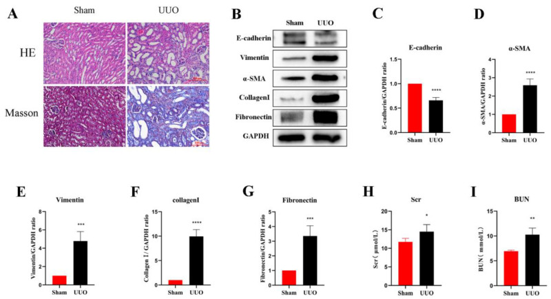
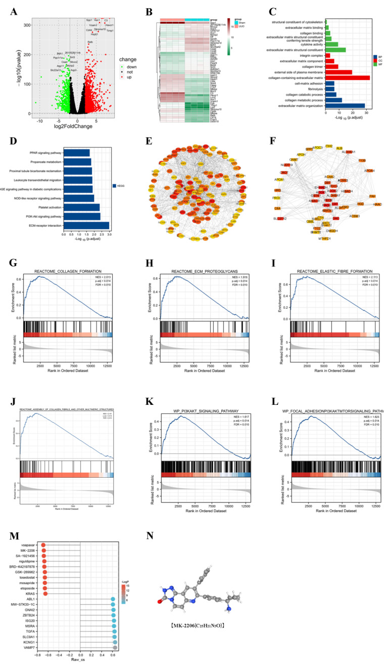
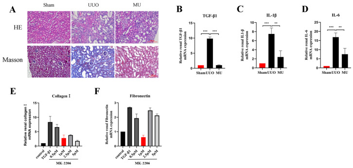
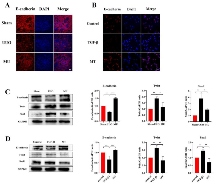
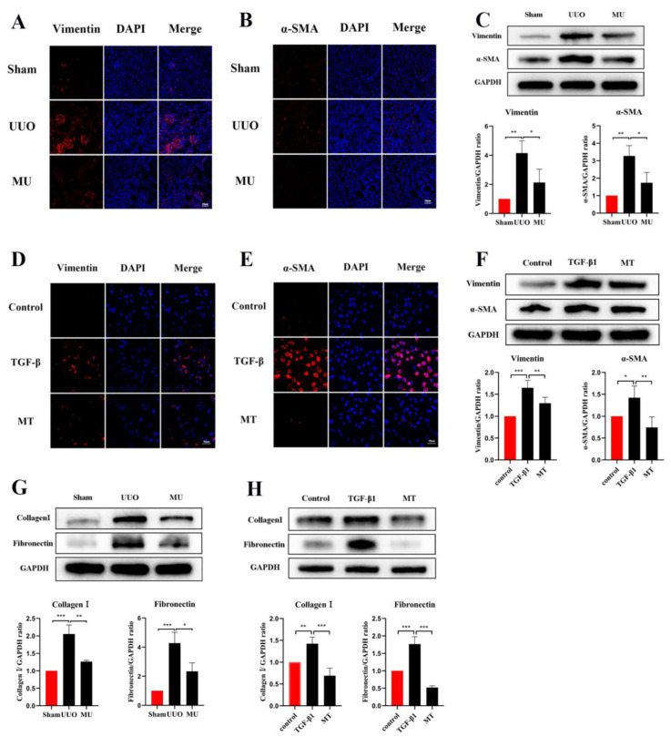
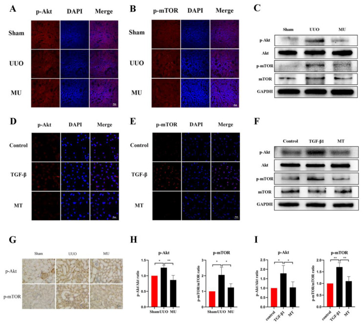
Renal fibrosis is a common pathological feature of various kidney diseases, leading to irreversible renal failure and end-stage renal disease. However, there are still no effective treatments to reverse renal fibrosis. This study aimed to explore the potential mechanism of a targeted drug for fibrosis. Here, unilateral ureteral obstruction (UUO)-treated mice and a TGF-β1-treated human renal tubular epithelial cell line (HK-2 cells) were used as models of renal fibrosis. Based on the changes of mRNA in UUO kidneys detected by transcriptome sequencing, MK-2206, an Akt inhibitor, was predicted as a potential drug to alleviate renal fibrosis through bioinformatics. We dissolved UUO mice with MK-2206 by gastric gavage and cultured TGF-β-induced HK-2 cells with MK-2206. Histopathological examinations were performed after MK-2206 intervention, and the degree of renal fibrosis, as well as the expression of Akt/mTOR pathway-related proteins, were evaluated by immunohistochemical staining, immunofluorescence staining, and Western blot. The results showed that MK-2206 significantly improved the pathological structure of the kidney. Furthermore, MK-2206 intervention effectively inhibited UUO- and TGF-β1-induced epithelial-mesenchymal transition, fibroblast activation, and extracellular matrix deposition. Mechanistically, MK-2206 treatment attenuated the activation of the Akt/mTOR signaling pathway. Taken together, our study revealed for the first time that MK-2206 is a promising drug for the improvement of renal fibrosis.
Keywords: Akt/mTOR signaling pathway; MK-2206; chronic kidney diseases; renal fibrosis.
Conflict of interest statement
The authors declare no conflict of interest.
Figures
Similar articles
-
Ruxolitinib Alleviates Renal Interstitial Fibrosis in UUO Mice.Int J Biol Sci. 2020 Jan 1;16(2):194-203. doi: 10.7150/ijbs.39024. eCollection 2020. Int J Biol Sci. 2020. PMID: 31929748 Free PMC article.
-
Aloe-Emodin Ameliorates Renal Fibrosis Via Inhibiting PI3K/Akt/mTOR Signaling Pathway In Vivo and In Vitro.Rejuvenation Res. 2019 Jun;22(3):218-229. doi: 10.1089/rej.2018.2104. Epub 2018 Oct 24. Rejuvenation Res. 2019. PMID: 30215298
-
Dedicator of cytokinesis protein 2 activates the epithelial-mesenchymal transition in renal fibrosis through the Rac1/PI3K/AKT pathway.Biochim Biophys Acta Mol Cell Res. 2025 Feb;1872(2):119894. doi: 10.1016/j.bbamcr.2024.119894. Epub 2024 Dec 24. Biochim Biophys Acta Mol Cell Res. 2025. PMID: 39725220
-
Hydroxychloroquine alleviates renal interstitial fibrosis by inhibiting the PI3K/Akt signaling pathway.Biochem Biophys Res Commun. 2022 Jun 25;610:154-161. doi: 10.1016/j.bbrc.2022.04.058. Epub 2022 Apr 15. Biochem Biophys Res Commun. 2022. PMID: 35462097
-
Nrf2 signaling attenuates epithelial-to-mesenchymal transition and renal interstitial fibrosis via PI3K/Akt signaling pathways.Exp Mol Pathol. 2019 Dec;111:104296. doi: 10.1016/j.yexmp.2019.104296. Epub 2019 Aug 23. Exp Mol Pathol. 2019. PMID: 31449784
Cited by
-
Inhibition of the NF-κB Signaling Pathway Alleviates Pyroptosis in Bladder Epithelial Cells and Neurogenic Bladder Fibrosis.Int J Mol Sci. 2023 Jul 6;24(13):11160. doi: 10.3390/ijms241311160. Int J Mol Sci. 2023. PMID: 37446339 Free PMC article.
-
Tubular FoxP2 and Kidney Fibrosis.J Am Soc Nephrol. 2025 Apr 1;36(4):544-558. doi: 10.1681/ASN.0000000576. Epub 2024 Dec 10. J Am Soc Nephrol. 2025. PMID: 39656554
-
Integrated oral microgel system ameliorates renal fibrosis by hitchhiking co-delivery and targeted gut flora modulation.J Nanobiotechnology. 2024 Jun 1;22(1):305. doi: 10.1186/s12951-024-02586-2. J Nanobiotechnology. 2024. PMID: 38822364 Free PMC article.
-
Suture-anchored cutaneous tension induces persistent hypertrophic scarring in a novel murine model.Burns Trauma. 2024 Oct 20;12:tkae051. doi: 10.1093/burnst/tkae051. eCollection 2024. Burns Trauma. 2024. PMID: 39429643 Free PMC article.
-
m6A eraser FTO impairs gemcitabine resistance in pancreatic cancer through influencing NEDD4 mRNA stability by regulating the PTEN/PI3K/AKT pathway.J Exp Clin Cancer Res. 2023 Aug 22;42(1):217. doi: 10.1186/s13046-023-02792-0. J Exp Clin Cancer Res. 2023. PMID: 37605223 Free PMC article.
References
-
- Bikbov B., Purcell C.A., Levey A.S., Smith M., Abdoli A., Abebe M., Adebayo O.M., Afarideh M., Agarwal S.K., Agudelo-Botero M., et al. Global, regional, and national burden of chronic kidney disease, 1990-2017: A systematic analysis for the Global Burden of Disease Study 2017. Lancet. 2020;395:709–733. doi: 10.1016/S0140-6736(20)30045-3. - DOI - PMC - PubMed
Publication types
MeSH terms
Substances
LinkOut - more resources
Full Text Sources
Medical
Miscellaneous
