. 2022 Nov 6;11(21):3512.
doi: 10.3390/cells11213512.

A Balance between Transmembrane-Mediated ER/Golgi Retention and Forward Trafficking Signals in Glycophorin-Anion Exchanger-1 Interaction

Affiliations

A Balance between Transmembrane-Mediated ER/Golgi Retention and Forward Trafficking Signals in Glycophorin-Anion Exchanger-1 Interaction

Kate Hsu et al. Cells. .

Abstract

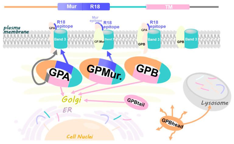
Anion exchanger-1 (AE1) is the main erythroid Cl-/HCO3- transporter that supports CO2 transport. Glycophorin A (GPA), a component of the AE1 complexes, facilitates AE1 expression and anion transport, but Glycophorin B (GPB) does not. Here, we dissected the structural components of GPA/GPB involved in glycophorin-AE1 trafficking by comparing them with three GPB variants-GPBhead (lacking the transmembrane domain [TMD]), GPBtail (mainly the TMD), and GP.Mur (glycophorin B-A-B hybrid). GPB-derived GP.Mur bears an O-glycopeptide that encompasses the R18 epitope, which is present in GPA but not GPB. By flow cytometry, AE1 expression in the control erythrocytes increased with the GPA-R18 expression; GYP.Mur+/+ erythrocytes bearing both GP.Mur and GPA expressed more R18 epitopes and more AE1 proteins. In contrast, heterologously expressed GPBtail and GPB were predominantly localized in the Golgi apparatus of HEK-293 cells, whereas GBhead was diffuse throughout the cytosol, suggesting that glycophorin transmembrane encoded an ER/Golgi retention signal. AE1 coexpression could reduce the ER/Golgi retention of GPB, but not of GPBtail or GPBhead. Thus, there are forward-trafficking and transmembrane-driven ER/Golgi retention signals encoded in the glycophorin sequences. How the balance between these opposite trafficking signals could affect glycophorin sorting into AE1 complexes and influence erythroid anion transport remains to be explored.

Keywords: ER/Golgi retention; GP.Mur (Miltenberger subtype III; Mi.III); SLC4A1); anion exchanger-1 (AE1; band 3; erythrocytes); glycophorin A (GPA); glycophorin B (GPB); membrane protein; oligomerization; red blood cells (RBCs; trafficking; transmembrane domain (TMD).


Conflict of interest statement

The authors declare no conflict of interest.

Figures

Figure 1
Figure 1
Structural-functional correlates for GPA/GPB and their variants. (A) Sequence alignments of GPA, GPB, and variants—GP.Mur, GPBtail, and GPBhead. Most C-terminal, cytoplasmic residues of GPA (25 amino acids) were omitted. The cytoplasmic region of GPA has been reported to support AE1 forward trafficking [33]. The pre-TMD of GPA (residues 61–70) supports the anion transport activity of AE1 [12,33]. GP.Mur is distinguished from GPB by an additional O-glycopeptide that presents the Mur and R18 epitopes; this additional extracellular sequence supports biosynthesis and surface expression of AE1 complexes [11]. This study identified an ER/Golgi retention signal encoded in the homologous glycophorin TM domain, which could be counteracted by the forward-trafficking signals encoded in cytoplasmic GPA and the R18 epitope, or by AE1 coexpression. (B) The cartoon depicts GPA/GPB and their variant GP.Mur in dimeric and tetrameric AE1 complexes (top view on the RBC membrane). Though AE1 can form dimers and tetramers and traffic to the plasma membrane without the assistance of GPA, GPA (yellow symbols) and AE1 (blue-green balls) generally interact in a one-to-one fashion. GPB (red symbols) is also present in erythroid AE1-associated complexes, and has been hypothesized to interact with AE1 indirectly through GPA. The copy numbers of GP.Mur (orange symbols) and GPB are about one-fifth the number of GPA on the erythrocyte membrane. Like GPA, GP.Mur directly binds AE1 and promotes AE1 surface expression.
Figure 2
Figure 2
AE1 levels were directly proportional to the GPA-R18 levels on the erythrocyte membrane; GYP.Mur+/+ RBCs generally expressed more AE1 and more R18 epitopes from both GPA and GP.Mur. Fresh RBC samples were immunostained with R18 mAb or with a mix of anti-AE1 mAbs BRIC 6 and BRIC 71 (1:500 dilution each), followed by secondary antibody labeling and flow cytometry. Here, each dot represents the relative levels of AE1 and the R18 epitope of a RBC sample. The geometric means for all non-Miltenberger RBC samples were averaged and set as 100%; all data were expressed in percentage (%). The linearly fitted line showed a rough correlation between the relative levels of AE1 and the R18 epitope on the erythrocyte surface. The data from homologous Mi.III (GYP.Mur+/+) samples were shown in crossed symbols.
Figure 3
Figure 3
Confocal images revealed subcellular localization of (A) singly expressed AE1, GPA, GPMURgfp, and GPBgfp; (BD) AE1 coexpressed with GPA, GPMURgfp, or GPBgfp in equimolar ratio; and (E) AE1 coexpressed with both GPA and GPBgfp in HEK-293 cells. GPBgfp and GPMURgfp fusion proteins were used for tracking, as there is no GPB- or GP.Mur-specific antibody available for immunolabeling. The transfected cells were fixed and permeabilized for immunostaining of surface and intracellular AE1 and GPA. To visualize AE1 expression, the cells were immunostained with a mix of mouse anti-AE1 mAbs (BRIC 6, BRIC 71, and BRIC170), followed by Alexa Fluor 568 or 647-conjugated anti-mouse antibody. To visualize GPA expression, the transfected cells were immunostained with Alexa Fluor 568-conjugated anti-GPA mAb BRIC163 that targets cytoplasmic GPA (absent in GPB/GP.Mur). In (E), HEK-293 cells were transfected with pcAE1, pcGPA and pGPBgfp (their plasmid molar ratio as 1:1:0.2 to mimic the relative levels in human erythrocytes). AE1 and GPA were immunostained as above. Their fluorescence signals were shown in pseudocolor: blue for GPA, red for AE1, and GPBgfp emitting green fluorescence. Scale bar = 25 μm.
Figure 3
Figure 3
Confocal images revealed subcellular localization of (A) singly expressed AE1, GPA, GPMURgfp, and GPBgfp; (BD) AE1 coexpressed with GPA, GPMURgfp, or GPBgfp in equimolar ratio; and (E) AE1 coexpressed with both GPA and GPBgfp in HEK-293 cells. GPBgfp and GPMURgfp fusion proteins were used for tracking, as there is no GPB- or GP.Mur-specific antibody available for immunolabeling. The transfected cells were fixed and permeabilized for immunostaining of surface and intracellular AE1 and GPA. To visualize AE1 expression, the cells were immunostained with a mix of mouse anti-AE1 mAbs (BRIC 6, BRIC 71, and BRIC170), followed by Alexa Fluor 568 or 647-conjugated anti-mouse antibody. To visualize GPA expression, the transfected cells were immunostained with Alexa Fluor 568-conjugated anti-GPA mAb BRIC163 that targets cytoplasmic GPA (absent in GPB/GP.Mur). In (E), HEK-293 cells were transfected with pcAE1, pcGPA and pGPBgfp (their plasmid molar ratio as 1:1:0.2 to mimic the relative levels in human erythrocytes). AE1 and GPA were immunostained as above. Their fluorescence signals were shown in pseudocolor: blue for GPA, red for AE1, and GPBgfp emitting green fluorescence. Scale bar = 25 μm.
Figure 4
Figure 4
Different from GPA and GP.Mur with substantial surface expression, singly expressed GPB was much retained in the ER but could be driven to the cell surface upon AE1 coexpression. (A) TOP: Expression of the fluorescent fusion GPAyfp alone (left), GPMURgfp alone (middle), and GPBgfp alone (right). BOTTOM: Overlay of each glycophorin-fluorescent fusion protein with the blue-fluorescent ER tracker. (B) HEK-293 cells cotransfected with equimolar pGPAyfp and pcAE1 were fixed and permeabilized on the second day post-transfection for immunofluorescence labeling of AE1 in red (mixed BRIC 6 + BRIC 71 + BRIC 170 mAb [1:500 dilution each] followed by anti-mouse IgG conjugated Alexa fluor 568 [1:200 dilution]). (C) HEK-293 cells were cotransfected with equimolar pGPMURgfp and pcAE1, followed by the same staining protocol as in (B). (D) HEK-293 cells were cotransfected with equimolar pGPBgfp and pcAE1, followed by the same staining protocol as in (B). In (BD), the left panel showed expression of the glycophorin-fluorescence fusion protein (in green color) upon AE1 coexpression; the middle panel showed expression of AE1 (in red color) upon glycophorin coexpression; the right panel showed the merged fluorescent signals of the glycophorin fusion (green), AE1 (red) and the ER tracker (blue).
Figure 5
Figure 5
GPB and GPBtail, but not GP.Mur or GPBhead, expressed predominantly in the Golgi apparatus, suggesting a Golgi retention signal embedded in the glycophorin TM domain. HEK-293 cells were transfected with only (A) GPMURgfp, (B) GPBgfp, (C) GPBhead-GFP, or (D) GPBtail-GFP. On the second day post-transfection, the cells were labeled with a lipid marker for the Golgi apparatus—BODIPY TR-ceramide (left panel), or with mouse anti-LAMP-1 (a protein marker for lysosomes) following AF568 conjugated anti-mouse antibody (right panel). Scale bar = 25 μm.
Figure 6
Figure 6
Subcellular localization of GBhead-GFP or GBtail-GFP was not affected by AE1 coexpression, suggesting that there was not even remote interaction between AE1 and the partial GPB structure. (TOP) HEK-293 cells were cotransfected with equal copy number of pGPBhead-GFP and pcAE1 plasmids, followed by immunolabeling with mouse anti-AE1 and then AF568 conjugated anti-mouse mAb. Similar to the GPBhead-GFP expression alone (Figure 5C), the distribution of GPBhead-GFP in AE1-coexpressed cells remained largely cytosolic and did not show any association with AE1 on the cell membrane. (BOTTOM) HEK-293 cells were cotransfected with equimolar pcAE1 and pGPBtail-GFP plasmids, followed by the same immunolabeling protocol to track AE1, as described above. Scale bar = 25 μm.
Figure 7
Figure 7
A working model illustrates different trafficking signals encoded in the glycophorin sequence and manifested in glycophorin-AE1 interaction. (TOP) The common sequence of GPA/GPB/GP.Mur and variants are divided in color-coded segments (from left to right, or the N- to the C-terminus): the N-terminal sequence (orange), the Mur epitope (light purple), the R18 epitope (purple); the pre-TMD region that interacts with band 3 and facilitates band 3-mediated anion transport (green); the TMD (pink), and the long C-terminal sequence only in GPA (dark gray). The 5 glycophorin variants studied are each presented in a hoop ring with color segments corresponding to the sequence segments described above. GPA contains all the sequence segments but the Mur epitope. GPB lacks the Mur and the R18 epitopes, as well as the long C-terminus unique to GPA. GP.Mur, evolved from GPB, contains the Mur and the R18 antigens, and also lacks the long C-terminus. GPBtail mainly expresses the TMD. GPBhead mainly expresses the N-terminal glycophorin sequence. The lack of the TMD in GPBhead makes it a diffusive, cytosolic protein destined for the lysosomes. Glycophorin transmembrane is essential for protein retention in the ER and the Golgi apparatus. The glycophorin-R18 epitope (purple) is adjacent to the pre-transmembrane region (green) that includes the Wrb motif (arisen from the AE1-GPA/GP.Mur interaction interface) and functions to assist band 3-mediated anion transport [12,24,26,33,40]. Thus, R18-mediated forward trafficking in GPA and GP.Mur may involve protein–protein interaction with band 3 and co-migration to the cell surface. The long C-terminal sequence of GPA also encodes a forward trafficking signal [33].

Similar articles

Cited by

References

    1. Cartron J.P., Rahuel C. Human erythrocyte glycophorins: Protein and gene structure analyses. Transfus. Med. Rev. 1992;6:63–92. doi: 10.1016/S0887-7963(92)70158-8. - DOI - PubMed
    1. Steck T.L. The band 3 protein of the human red cell membrane: A review. J. Supramol. Struct. 1978;8:311–324. doi: 10.1002/jss.400080309. - DOI - PubMed
    1. Steck T.L., Ramos B., Strapazon E. Proteolytic dissection of band 3, the predominant transmembrane polypeptide of the human erythrocyte membrane. Biochemistry. 1976;15:1153–1161. doi: 10.1021/bi00650a030. - DOI - PubMed
    1. Knauf P.A., Gasbjerg P.K., Brahm J. The asymmetry of chloride transport at 38 degrees C in human red blood cell membranes. J. Gen. Physiol. 1996;108:577–589. doi: 10.1085/jgp.108.6.577. - DOI - PMC - PubMed
    1. Alper S.L. The band 3-related anion exchanger (AE) gene family. Annu Rev. Physiol. 1991;53:549–564. doi: 10.1146/annurev.ph.53.030191.003001. - DOI - PubMed

Publication types

LinkOut - more resources