. 2022 Nov 7;11(21):3515.
doi: 10.3390/cells11213515.

PI3K Isoform-Specific Regulation of Leader and Follower Cell Function for Collective Migration and Proliferation in Response to Injury

Affiliations

PI3K Isoform-Specific Regulation of Leader and Follower Cell Function for Collective Migration and Proliferation in Response to Injury

Morgan D Basta et al. Cells. .

Abstract

To ensure proper wound healing it is important to elucidate the signaling cues that coordinate leader and follower cell behavior to promote collective migration and proliferation for wound healing in response to injury. Using an ex vivo post-cataract surgery wound healing model we investigated the role of class I phosphatidylinositol-3-kinase (PI3K) isoforms in this process. Our findings revealed a specific role for p110α signaling independent of Akt for promoting the collective migration and proliferation of the epithelium for wound closure. In addition, we found an important role for p110α signaling in orchestrating proper polarized cytoskeletal organization within both leader and wounded epithelial follower cells to coordinate their function for wound healing. p110α was necessary to signal the formation and persistence of vimentin rich-lamellipodia extensions by leader cells and the reorganization of actomyosin into stress fibers along the basal domains of the wounded lens epithelial follower cells for movement. Together, our study reveals a critical role for p110α in the collective migration of an epithelium in response to wounding.

Keywords: PI3K; collective migration; follower cell; leader cell; p110α; wound healing.


Conflict of interest statement

The authors declare no conflict of interest.

Figures
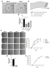
Figure 1
Figure 1
Wound healing in response to cataract surgery injury occurs in a PI3K isoform-specific manner. (A) Depiction of ex vivo post-cataract surgery culture to study mechanisms of wound repair across the cell-denuded endogenous basement membrane in the center of the lens capsule. Ex vivo post-cataract surgery explant on day 0 depicts the wounded lens epithelial cells within the original attachment zone (OAZ) and the cell-denuded central migration zone (CMZ) (i). In response to injury, collective migration across the endogenous basement membrane in the CMZ is driven by leader cells that direct the migration of the wounded epithelial follower cells (ii), arrow depicts the direction of wound healing. Collective migration across the CMZ for wound repair is completed by day 3 post-injury (iii). (B,C) Ex vivo post-cataract surgery explants were treated from Time 0 through Day 3 with individual PI3K isoform inhibitors, HS-173 for p110α; GSK2636771 for p110β; or Duvelisib for p110δ/γ. (B) Graph from Wes analysis depicts the efficacy of individual PI3K isoform inhibitors compared to vehicle control by examining the relative intensity of the PI3K effector, phosphorylated-AKT (pAKT) relative to total AKT expression compared to GAPDH (loading control). (C) Phase microscopy shows open wound area (dotted black circle) for vehicle vs. PI3K inhibitor treatment from Day 0 through Day 3. Graph depicts % area of wound closure. Results show that blocking p110α with HS-173 had the largest impact on inhibiting wound closure. (D,E) Ex vivo post-cataract surgery explants were treated with vehicle (DMSO) or the AKT specific inhibitor, MK-2206 from Time 0 through Day 3. (D) MK-2206 suppressed pAKT relative to total AKT expression and GAPDH (loading control). (E) Graph of % wound closure shows that treatment with MK-2206 had no effect on wound closure. p-values were determined from one-way ANOVA (B), two-way ANOVA with multiple comparisons (B,C) or unpaired t-test (D,E). (B, * p < 0.05, ** p < 0.01; C, **** p < 0.0001 and D **** p < 0.0001). Data is expressed as ± SEM from 2 (D,E) or 3 (B,C) independent experiments. Magnification bars = 1000 μm (A,C).
Figure 2
Figure 2
p110α is required to initiate extension of vimentin rich lamellipodial of leader cells. (A) Model of CMZ highlighting our region of interest (leader cells, black circle) for these studies. (BG) Ex vivo post-cataract surgery explants were treated with HS-173 to inhibit p110α from Time 0 for 24 h (Day 1 post-injury). (B) Phase contrast imaging shows that in the presence of DMSO leader cells extend protrusions at the wound edge. (D) In contrast, HS-173 blocked leader cell protrusion at the wound edge. Regions in (B,D) are shown at higher magnification (C,E). (F,G) Cataract surgery explants day 1 post-treatment were immunolabeled for vimentin (green) and counterstained for DAPI (blue). Confocal images revealed vimentin-rich lamellipodia extensions at the leading edge were blocked by HS-173 treatment (G) compared to vehicle control (F). Magnification bars = 50 μm (BE) and 20 μm (F,G).
Figure 3
Figure 3
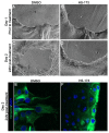
p110α is required to maintain the extension of vimentin-rich lamellipodia of leader cells. (AF) Ex vivo post-cataract surgery explants were treated with vehicle (DMSO) or HS-173 to inhibit p110α from Day 1 for 24 h (Day 2 post-injury). (A,B) Phase contrast imaging on Day 1 shows the extension of protrusions by leader cells at the wound edge prior to treatment. (C,D) In contrast, phase contrast imaging on Day 2 (24 h post-treatment) revealed that HS-173-treated leader cells failed to maintain protrusions compared to vehicle controls. (E,F) Day 2 explants following 24 h of treatment were immunolabeled for vimentin (green) and counterstained for DAPI (blue). Confocal images revealed that maintenance of vimentin-rich lamellipodia extensions was blocked by HS-173 treatment (F) compared to vehicle controls (E). Magnification bars = 50 μm (AD) and 20 μm (E,F).
Figure 4
Figure 4
p110α is required throughout the wound healing process to sustain wound closure. Ex vivo post-cataract surgery explants were treated with HS-173 to inhibit p110α from Day 1 to Day 3 post-injury. Phase microscopy and wound area measurements show the wound area prior to treatment on Day 0 and Day 1 and post-treatment with vehicle or HS-173 on Day 2 and Day 3 (dotted black circle). Graph depicts % wound closure and revealed that HS-173 is required throughout the wound healing process to drive wound closure. p-values were determined from two-way ANOVA with multiple comparisons (** p < 0.01). Data is expressed as ±SEM from 3 independent experiments. Magnification bars = 1000 μm.
Figure 5
Figure 5
Impact of blocking p110α on the apical organization of the wounded lens epithelial follower cells post-injury. (A) Model of CMZ highlighting our region of interest (follower cells, black circle) for these studies. (BM) Ex vivo post-cataract surgery explants were treated with vehicle control or HS-173 to inhibit p110α from Time 0 for 24 h (Day 1 post-injury). Cultures were labeled with phalloidin to identify filamentous actin (F-actin, white (B,E), purple (D,G)) and immunolabeled for active myosin (p-myosin, white (C,F), green (D,G)). (BG) Confocal images of follower cells focused apically revealed a cortical distribution of actomyosin at cell–cell border with vehicle and HS-173 treatment. However, HS-173 treatment revealed differences in active myosin distribution at apical vertices (see arrowheads) and a more compact organization of the monolayer compared to vehicle control (E,G). 3D structures from confocal z-stacks, which were labeled for F-actin (white) and nuclei (DAPI, blue) were created to allow the better visualization of the compact organization of wounded epithelial follower cells with HS-173 and vehicle treatment (H,I). (J,K) Orthogonal views from confocal z-stacks of cultures labeled for F-actin (white) and nuclei (DAPI, blue) were created to reveal height differences in the wounded epithelial cells of HS-173 vs. DMSO-treated cultures. (L,M) 3D structures of orthogonal views were created from the same region in (J,K) to further highlight shape differences. Magnification bars = 20 μm (BG,J,K) or 5 μm (H,I). (BG) arrows show direction of cell migration.
Figure 6
Figure 6
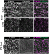
Role for p110α in regulating reorganization of actomyosin along the basal surface of the lens epithelial follower cells for wound healing. (AF) Ex vivo post-cataract surgery explants were treated with vehicle control or HS-173 to inhibit p110α from Time 0 for 24 h (Day 1 post-injury). Cultures were labeled with phalloidin to identify filamentous actin (F-actin, white (A,D,G), purple (C,F,I)) and immunolabeled for active myosin II (P-myosin, white (B,E,H), green (C,F,I)). Confocal images along the basal surface of the wounded lens epithelium revealed that (AC) normal basal actomyosin reorganization into stress fibers within wounded epithelial cells is blocked with (DF) HS-173 treatment. (GI) Confocal images along the basal surface of the wounded epithelial cell located in the OAZ are shown for comparison to HS-173 treated cells (DF) revealing a similarity in basal actomyosin organization of HS-173 basal follower cells to nonmoving wounded epithelial cells in the OAZ. Magnification bars = 20 μm and arrows show direction of cell migration (AI).
Figure 7
Figure 7
p110α is required for promoting cell proliferation in the central migratory zone during wound healing. (AE) Time 0 ex vivo post-cataract surgery explants were treated with vehicle (DMSO) or HS-173 to inhibit p110α for 24 h (Day 1 post-injury) and labeled for 30min with EdU (green) (A,C) and counterstained with DAPI (blue) (B,D). (E) Graph depicts the change in the relative % of EdU positive cells within the CMZ region in vehicle compared to HS-173 treated cultures. Cell proliferation was blocked with HS-173 treatment. Vehicle was normalized to 100%. p-values were determined from unpaired t-test (E, **** p < 0.0001). Data is expressed as ± SEM from 3 independent experiments (E). Magnification bars = 20 μm (AD).

References

    1. Jain S., Ladoux B., Mege R.M. Mechanical plasticity in collective cell migration. Curr. Opin. Cell Biol. 2021;72:54–62. doi: 10.1016/j.ceb.2021.04.006. - DOI - PubMed
    1. Qin L., Yang D., Yi W., Cao H., Xiao G. Roles of leader and follower cells in collective cell migration. Mol. Biol. Cell. 2021;32:1267–1272. doi: 10.1091/mbc.E20-10-0681. - DOI - PMC - PubMed
    1. Rorth P. Collective cell migration. Annu. Rev. Cell Dev. Biol. 2009;25:407–429. doi: 10.1146/annurev.cellbio.042308.113231. - DOI - PubMed
    1. Capuana L., Bostrom A., Etienne-Manneville S. Multicellular scale front-to-rear polarity in collective migration. Curr. Opin. Cell Biol. 2020;62:114–122. doi: 10.1016/j.ceb.2019.10.001. - DOI - PubMed
    1. Mayor R., Etienne-Manneville S. The front and rear of collective cell migration. Nat. Rev. Mol. Cell Biol. 2016;17:97–109. doi: 10.1038/nrm.2015.14. - DOI - PubMed

Publication types

Substances

LinkOut - more resources