. 2022 Nov 7;11(21):3523.
doi: 10.3390/cells11213523.

Branched-Chain Amino Acids Are Linked with Alzheimer's Disease-Related Pathology and Cognitive Deficits

Affiliations

Branched-Chain Amino Acids Are Linked with Alzheimer's Disease-Related Pathology and Cognitive Deficits

Md Abu Bakkar Siddik et al. Cells. .

Abstract

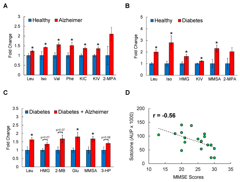
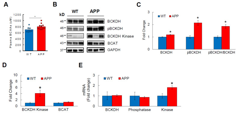
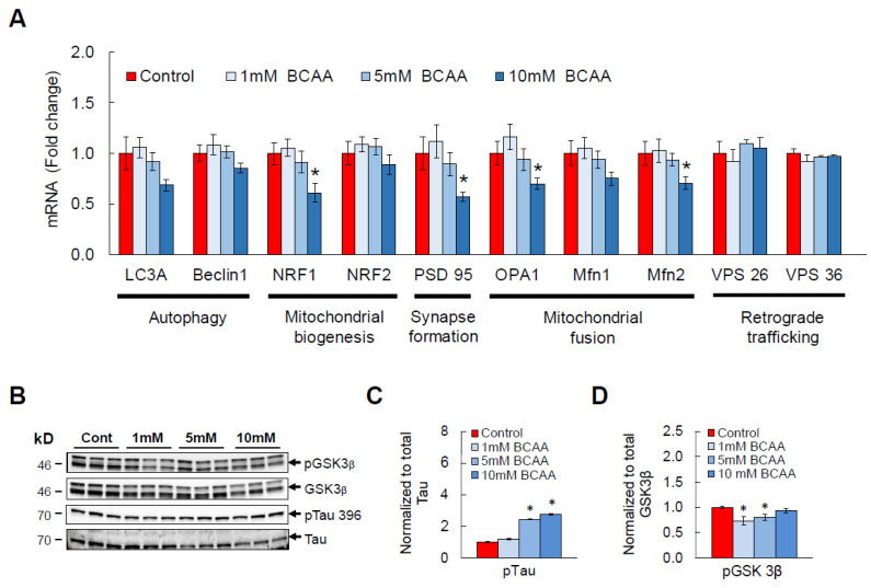
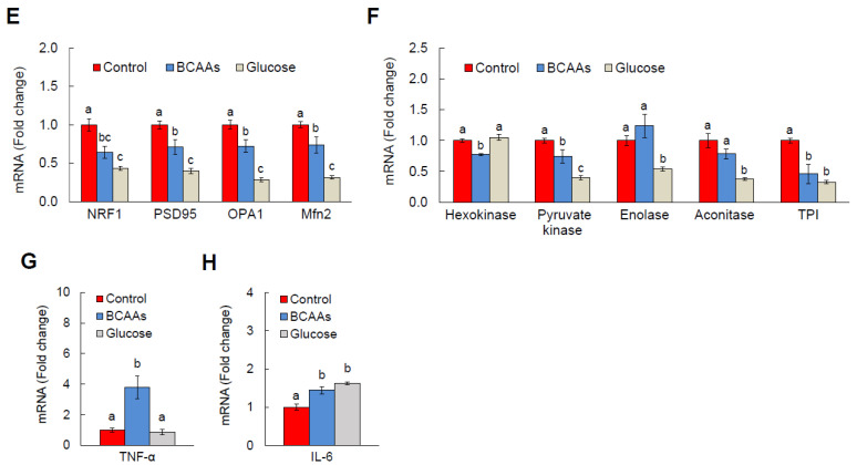
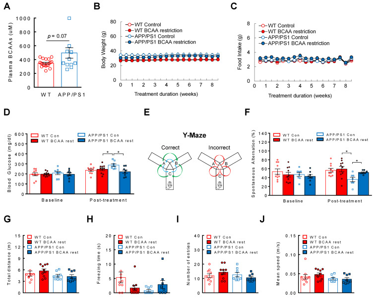
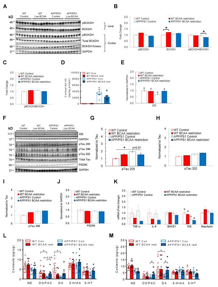
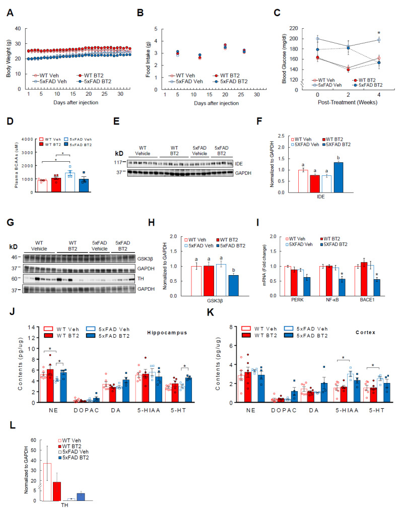
Alzheimer's disease (AD) is an irreversible neurodegenerative disorder with a complex pathophysiology. Type 2 diabetes (T2D) is a strong risk factor for AD that shares similar abnormal features including metabolic dysregulation and brain pathology such as amyloid and/or Tau deposits. Emerging evidence suggests that circulating branched-chain amino acids (BCAAs) are associated with T2D. While excess BCAAs are shown to be harmful to neurons, its connection to AD is poorly understood. Here we show that individuals with AD have elevated circulating BCAAs and their metabolites compared to healthy individuals, and that a BCAA metabolite is correlated with the severity of dementia. APPSwe mouse model of AD also displayed higher plasma BCAAs compared to controls. In pursuit of understanding a potential causality, BCAA supplementation to HT-22 neurons was found to reduce genes critical for neuronal health while increasing phosphorylated Tau. Moreover, restricting BCAAs from diet delayed cognitive decline and lowered AD-related pathology in the cortex and hippocampus in APP/PS1 mice. BCAA restriction for two months was sufficient to correct glycemic control and increased/restored dopamine that were severely reduced in APP/PS1 controls. Treating 5xFAD mice that show early brain pathology with a BCAA-lowering compound recapitulated the beneficial effects of BCAA restriction on brain pathology and neurotransmitters including norepinephrine and serotonin. Collectively, this study reveals a positive association between circulating BCAAs and AD. Our findings suggest that BCAAs impair neuronal functions whereas BCAA-lowering alleviates AD-related pathology and cognitive decline, thus establishing a potential causal link between BCAAs and AD progression.

Keywords: 5xFAD; APP/PS1; Aβ-42; BCAA; Tau; diet restriction; glucose metabolism; neurotransmitters.


Conflict of interest statement

The authors declare no conflict of interest.

Figures

Figure 1
Figure 1
Circulating BCAA levels and their metabolites are elevated in individuals with AD. Serum samples from age- and BMI-matched healthy controls, patients with AD, T2D, or with T2D+AD after overnight fasting were analyzed by LC/MS. (A) Healthy vs. AD; (B) Healthy vs. Diabetes; (C) Diabetes vs. Diabetes+AD. Data shown are fold changes normalized to the mean of healthy or Diabetes controls; n = 10 males/group; mean age = 72 ± 3 years old; * p < 0.05. (D) Correlation analysis between serum sotolone, a metabolite of isoleucine, and MMSE scores. Iso: Isoleucine; HMG: 3-methylglutaryl (metabolite of leucine); KIV: ketoisovalerate (metabolite of valine); KIC: ketoisocaproate (metabolite of leucine); MMSA: Methyl-melonatesemialdehyde (metabolite of valine); Phe: Phenylalanine; 2-MPA: 2-methylpropanoic acid (metabolite of valine); 3-HP: 3-OH-Propanoate (metabolite of valine); MMSE: Mini-Mental State Examination.
Figure 2
Figure 2
BCAA metabolism is impaired in transgenic APPSwe mice. 8-month-old WT or APPSwe AD mice were sacrificed after overnight fasting. (A) Fasting plasma BCAA levels measured by spectrophotometric assay. (B) Western blots showing liver protein abundance of BCKDH, the rate-limiting enzyme in BCAA breakdown; phosphorylated, inactive state of BCKDH (pBCKDH); BCKDH kinase, an enzyme that phosphorylates BCKDH; and BCAT, an enzyme involved in the reversible first step in BCAA degradation. (C) Analysis of BCKDH, pBCKDH, and the inactivity index (pBCKDH/BCKDH ratio). (D) Analysis of BCKDH kinase and BCAT in liver. (E) mRNA expression analysis of BCKDH, BCKDH phosphatase, and BCKDH kinase via RT-qPCR in liver normalized to GAPDH. n = 7–13 males/group; Analyzed by student’s t-test. * p < 0.05. Phosphatase—BCKDH phosphatase; Kinase—BCKDH kinase.
Figure 3
Figure 3
BCAA treatment induces AD-related changes in vitro. Differentiated HT-22 hippocampal neurons were supplemented with a mixture of BCAAs in the culture media for 24 h. (A) mRNA expression of genes involved in autophagy, mitochondrial biogenesis and fusion, synapse formation, and retrograde trafficking. (B) Western blots showing pGSK3β, total GSK3β, pTau 396, total Tau. (C) Analysis of Tau phosphorylation at residue 396 and (D) pGSK3β. (EH) In a separate cohort, differentiated HT-22 neurons were exposed to either vehicle or 10 mM BCAAs in the media for 24 h. Another group was exposed to 25 mM glucose to induce glucotoxicity, a widely used neurotoxicity model, as a comparison. (E) mRNA levels for neuronal health genes as described above were analyzed. (F) Genes critical for glycolytic pathway. (G) mRNAs of TNF-α and (H) IL-6. All mRNAs were normalized to B2M. n = 6/group; * p < 0.05 compared to Control. Groups with different letters (i.e., a, b, c) are significantly different from each other with p < 0.05.
Figure 3
Figure 3
BCAA treatment induces AD-related changes in vitro. Differentiated HT-22 hippocampal neurons were supplemented with a mixture of BCAAs in the culture media for 24 h. (A) mRNA expression of genes involved in autophagy, mitochondrial biogenesis and fusion, synapse formation, and retrograde trafficking. (B) Western blots showing pGSK3β, total GSK3β, pTau 396, total Tau. (C) Analysis of Tau phosphorylation at residue 396 and (D) pGSK3β. (EH) In a separate cohort, differentiated HT-22 neurons were exposed to either vehicle or 10 mM BCAAs in the media for 24 h. Another group was exposed to 25 mM glucose to induce glucotoxicity, a widely used neurotoxicity model, as a comparison. (E) mRNA levels for neuronal health genes as described above were analyzed. (F) Genes critical for glycolytic pathway. (G) mRNAs of TNF-α and (H) IL-6. All mRNAs were normalized to B2M. n = 6/group; * p < 0.05 compared to Control. Groups with different letters (i.e., a, b, c) are significantly different from each other with p < 0.05.
Figure 4
Figure 4
BCAA restriction delays the onset of cognitive deficit in APP/PS1 mice. 11-month-old WT or APP/PS1 mice without cognitive impairment were placed on a control diet or BCAA-restricted (50%) diet, iso-caloric and iso-nitrogenous, for two months. (A) Plasma BCAA levels at baseline. (B) BCAA restriction does not affect body weight or (C) Food intake. (D) Blood glucose before and after treatment. (E) Schematic of Y-maze behavioral test. (F) Spontaneous alternation (%) before and after treatment. (G) Total distance traveled, (H) Freezing time, (I) Number of arm entries, and (J) Mean speed during Y-maze test. WT control or BCAA restriction group (n = 10/group); APP/PS1 control or BCAA restriction group (n = 8/group). * p < 0.05.
Figure 5
Figure 5
BCAA restriction lowers brain pathology and restores neurotransmitter content in APP/PS1 mice. 11-month-old WT or APP/PS1 mice without cognitive impairment were placed on a control or BCAA-restricted (50%) diet that is iso-caloric and iso-nitrogenous for two months. (A) Western blots for proteins involved in BCAA degradation in liver and cortex of the brain. (B) pBCKDH and total BCKDH in liver at the end of two months. (C) BCKDH inactivity index (pBCKDH/BCKDH) in the cortex. (D) Cortical Aβ-42 levels normalized to protein by ELISA. (E) Insulin-degrading enzyme (IDE) in the cortex of the brain. (F) Western blots for IDE, PSD95, and phosphorylated Tau in the cortex. (G) Phosphorylated state of Tau at threonine residue 205, (H) Serine residue 202, and (I) Serine residue 396. (J) Protein expression of PSD95. (K) Analysis of genes involved in neuroinflammation and amyloid production and degradation, normalized to B2M. (L) Monoamine neurotransmitter concentrations in the hippocampus and (M) Cortex. NE—Norepinephrine; DA—Dopamine; DOPAC—Dopamine metabolite; 5-HT—Serotonin; 5-HIAA—Serotonin metabolite. * p < 0.05. Groups with different letters are significantly different from each other with p < 0.05.
Figure 6
Figure 6
BT2 is effective in alleviating brain pathology and increasing neurotransmitters in 5xFAD mice. WT or 5xFAD mice that are 6–8 weeks old were injected with either vehicle or BT2 (40 mg/kg/day) for one month. Animals were single-housed during the experiment. (A) Daily body weight. (B) Weekly food intake. (C) Blood glucose at baseline and at two and four weeks post-treatment. (D) Plasma BCAA levels at baseline and post-treatment. (E) Western blots for IDE in the cortex. (F) Protein analysis of IDE. (G) Western blots for GSK3β in the cortex and tyrosine hydroxylase (TH), the rate-limiting enzyme for catecholamine synthesis, in the hippocampus. (H) Protein expression of GSK3β. (I) Hippocampal mRNA abundance of markers for ER stress (PERK), inflammation (NF-κB), and amyloid synthesis (BACE1; also known as β-secretase), normalized to B2M. (J) Concentration of neurotransmitters (NE, DA, 5-HT) and their metabolites (DOPAC, 5-HIAA) measured in the hippocampus and (K) Cortex. (L) TH protein expression in the hippocampus. WT vehicle or BT2 group (n = 6/group); 5xFAD vehicle or BT2 group (n = 4/group). * p < 0.05. Groups with different letters are significantly different from each other with p < 0.05.

Similar articles

Cited by

References

    1. Alzheimer’s Association Alzheimer’s Disease Facts and Figures. [(accessed on 8 September 2022)]. Available online: https://www.alz.org/alzheimers-dementia/facts-figures.
    1. Salas I.H., De Strooper B. Diabetes and Alzheimer’s Disease: A Link not as Simple as it Seems. Neurochem. Res. 2019;44:1271–1278. doi: 10.1007/s11064-018-2690-9. - DOI - PubMed
    1. Umegaki H. Type 2 diabetes as a risk factor for cognitive impairment: Current insights. Clin. Interv. Aging. 2014;9:1011–1019. doi: 10.2147/CIA.S48926. - DOI - PMC - PubMed
    1. Adams S.H., Hoppel C.L., Lok K.H., Zhao L., Wong S.W., Minkler P.E., Hwang D.H., Newman J.W., Garvey W.T. Plasma acylcarnitine profiles suggest incomplete long-chain fatty acid beta-oxidation and altered tricarboxylic acid cycle activity in type 2 diabetic African-American women. J. Nutr. 2009;139:1073–1081. doi: 10.3945/jn.108.103754. - DOI - PMC - PubMed
    1. Mihalik S.J., Goodpaster B.H., Kelley D.E., Chace D.H., Vockley J., Toledo F.G., DeLany J.P. Increased levels of plasma acylcarnitines in obesity and type 2 diabetes and identification of a marker of glucolipotoxicity. Obesity. 2010;18:1695–1700. doi: 10.1038/oby.2009.510. - DOI - PMC - PubMed

Publication types

Substances

LinkOut - more resources