

Cytogenetic Investigation of Infertile Patients in Hungary: A 10-Year Retrospective Study
- PMID: 36360324
- PMCID: PMC9690888
- DOI: 10.3390/genes13112086
Cytogenetic Investigation of Infertile Patients in Hungary: A 10-Year Retrospective Study
Abstract
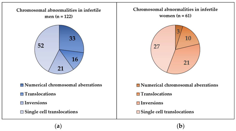
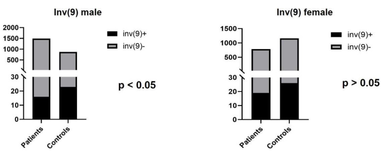
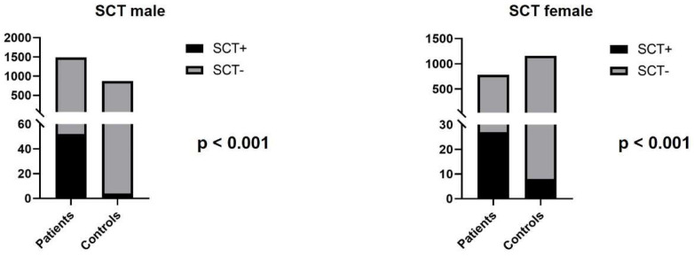
Chromosome abnormalities play a crucial role in reproductive failure. The presence of numerical or structural aberrations may induce recurrent pregnancy loss or primary infertility. The main purpose of our study was to determine the types and frequency of chromosomal aberrations in infertile patients and to compare the frequency of structural aberrations to a control group. Karyotyping was performed in 1489 men and 780 women diagnosed with reproductive failure between 2010 and 2020. The control group included 869 male and 1160 female patients having cytogenetic evaluations for reasons other than infertility. Sex chromosomal aberrations were detected in 33/1489 (2.22%) infertile men and 3/780 (0.38%) infertile women. Structural abnormalities (e.g., translocation, inversion) were observed in 89/1489 (5.98%) infertile men and 58/780 (7.44%) infertile women. The control population showed structural chromosomal abnormalities in 27/869 (3.11%) men and 39/1160 (3.36%) women. There were significant differences in the prevalence of single-cell translocations between infertile individuals (males: 3.5%; females: 3.46%) and control patients (males: 0.46%; females: 0.7%). In summary, this is the first report of cytogenetic alterations in infertile patients in Hungary. The types of chromosomal abnormalities were comparable to previously published data. The prevalence of less-studied single-cell translocations was significantly higher in infertile patients than in the control population, supporting an earlier suggestion that these aberrations may be causally related to infertility.
Keywords: chromosomal aberrations; cytogenetics; infertility; single-cell translocations.
Conflict of interest statement
The authors declare no conflict of interest.
Figures

Similar articles
-
Prevalence of Chromosomal Abnormalities in Iranian Patients with Infertility.Arch Iran Med. 2023 Feb 1;26(2):110-116. doi: 10.34172/aim.2023.17. Arch Iran Med. 2023. PMID: 37543931 Free PMC article.
-
Cytogenetic studies in patients with reproductive failure.Acta Obstet Gynecol Scand. 2003 Jan;82(1):53-6. doi: 10.1034/j.1600-0412.2003.820109.x. Acta Obstet Gynecol Scand. 2003. PMID: 12580840
-
Cytogenetic Abnormalities Found in Patients with Reproductive Problems.Med Arch. 2017 Dec;71(6):396-399. doi: 10.5455/medarh.2017.71.396-399. Med Arch. 2017. PMID: 29416198 Free PMC article.
-
[Value of karyotyping women patients of couples referred for sterility].Gynecol Obstet Fertil. 2003 Jan;31(1):66-9. doi: 10.1016/s1297-9589(02)00008-5. Gynecol Obstet Fertil. 2003. PMID: 12659787 Review. French.
-
[Benefit of human gamete cytogenetics: results and perspectives].Pathol Biol (Paris). 2008 Sep;56(6):388-99. doi: 10.1016/j.patbio.2008.04.012. Epub 2008 Jun 4. Pathol Biol (Paris). 2008. PMID: 18534785 Review. French.
Cited by
-
Fertility problems in men carrying chromosome 7 inversion: A retrospective, observational study.Medicine (Baltimore). 2025 Jan 17;104(3):e41358. doi: 10.1097/MD.0000000000041358. Medicine (Baltimore). 2025. PMID: 39833054 Free PMC article.
-
Genomic aspects in reproductive medicine.Clin Exp Reprod Med. 2024 Jun;51(2):91-101. doi: 10.5653/cerm.2023.06303. Epub 2024 Jan 24. Clin Exp Reprod Med. 2024. PMID: 38263590 Free PMC article.
-
Evaluation of chromosomal abnormalities in the postnatal cohort: A single-center study on 14,242 patients.J Clin Lab Anal. 2024 Jan;38(1-2):e24997. doi: 10.1002/jcla.24997. Epub 2023 Dec 19. J Clin Lab Anal. 2024. PMID: 38115218 Free PMC article.
References
MeSH terms
LinkOut - more resources
Full Text Sources
Medical
