

Advanced Endoscopy for Benign Esophageal Disease: A Review Focused on Non-Erosive Reflux Disease and Eosinophilic Esophagitis
- PMID: 36360524
- PMCID: PMC9690083
- DOI: 10.3390/healthcare10112183
Advanced Endoscopy for Benign Esophageal Disease: A Review Focused on Non-Erosive Reflux Disease and Eosinophilic Esophagitis
Abstract
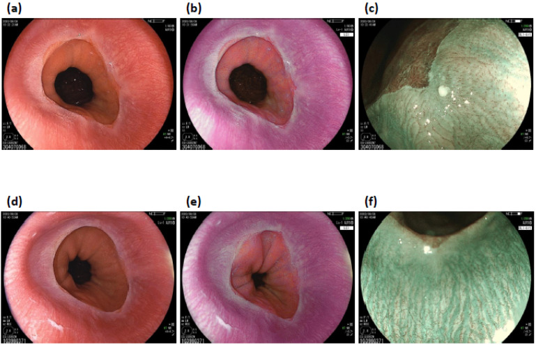
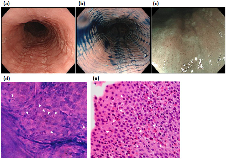
Advanced endoscopy (AVE) techniques include image-enhanced endoscopy methods, such as narrow-band imaging (NBI), and types of microscopic endoscopy, such as endocytoscopy. In the esophagus, AVE first showed diagnostic utility in the diagnosis of superficial esophageal cancer and was then applied to inflammatory disease. This review focuses on non-erosive reflux disease (NERD) and eosinophilic esophagitis (EoE), which sometimes show no abnormal findings on standard white light endoscopy alone. Studies have demonstrated that advanced endoscopy, including NBI magnification endoscopy and endocytoscopy, improved the diagnostic performance of white-light endoscopy alone for NERD and EoE. In this review, we explain why advanced endoscopy is needed for the diagnosis of these esophageal inflammatory diseases, summarize the study results, and discuss future perspectives.
Keywords: endocytoscopy; endoscopic diagnosis; eosinophilic esophagitis; esophagus; gastroesophageal reflux disease (GERD); image enhanced endoscopy; magnification endoscopy; narrow-band imaging; non-erosive reflux disease (NERD).
Conflict of interest statement
The authors declare no conflict of interest.
Figures
Similar articles
-
Correlation of narrow band imaging endoscopy and histopathology in the diagnosis of nonerosive reflux disease.Saudi J Gastroenterol. 2015 Sep-Oct;21(5):330-6. doi: 10.4103/1319-3767.164205. Saudi J Gastroenterol. 2015. PMID: 26458862 Free PMC article.
-
Endoscope versus microscope in the diagnosis of esophageal non-erosive reflux disease: a study of 71 cases.Malays J Pathol. 2014 Dec;36(3):181-8. Malays J Pathol. 2014. PMID: 25500517 Clinical Trial.
-
Autofluorescence imaging endoscopy can distinguish non-erosive reflux disease from functional heartburn: A pilot study.World J Gastroenterol. 2016 Apr 14;22(14):3845-51. doi: 10.3748/wjg.v22.i14.3845. World J Gastroenterol. 2016. PMID: 27076770 Free PMC article.
-
Potential contribution of novel imaging modalities in non-erosive reflux disease.Best Pract Res Clin Gastroenterol. 2008;22(4):617-24. doi: 10.1016/j.bpg.2008.03.001. Best Pract Res Clin Gastroenterol. 2008. PMID: 18656820 Review.
-
A critical assessment of the current status of non-erosive reflux disease.Digestion. 2008;78 Suppl 1:46-54. doi: 10.1159/000151255. Epub 2008 Oct 2. Digestion. 2008. PMID: 18832840 Review.
Cited by
-
Cyber-Physical System Interface for Implantable Esophageal Prosthesis.Sensors (Basel). 2025 Jul 18;25(14):4469. doi: 10.3390/s25144469. Sensors (Basel). 2025. PMID: 40732597 Free PMC article.
References
-
- Muto M., Minashi K., Yano T., Saito Y., Oda I., Nonaka S., Omori T., Sugiura H., Goda K., Kaise M., et al. Early detection of superficial squamous cell carcinoma in the head and neck region and esophagus by narrow band imaging: A multicenter randomized controlled trial. J. Clin. Oncol. 2010;28:1566–1572. doi: 10.1200/JCO.2009.25.4680. - DOI - PMC - PubMed
-
- Ezoe Y., Muto M., Uedo N., Doyama H., Yao K., Oda I., Kaneko K., Kawahara Y., Yokoi C., Sugiura Y., et al. Magnifying narrowband imaging is more accurate than conventional white-light imaging in diagnosis of gastric mucosal cancer. Gastroenterology. 2011;141:2017–2025.e3. doi: 10.1053/j.gastro.2011.08.007. - DOI - PubMed
Publication types
LinkOut - more resources
Full Text Sources
