. 2022 Oct 28;19(21):14088.
doi: 10.3390/ijerph192114088.

Oral Microcosm Biofilms Grown under Conditions Progressing from Peri-Implant Health, Peri-Implant Mucositis, and Peri-Implantitis

Affiliations

Oral Microcosm Biofilms Grown under Conditions Progressing from Peri-Implant Health, Peri-Implant Mucositis, and Peri-Implantitis

Vanessa Sousa et al. Int J Environ Res Public Health. .

Abstract

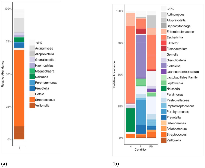
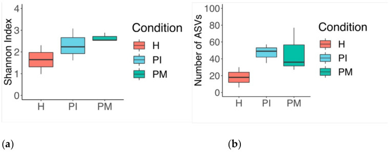
Peri-implantitis is a disease influenced by dysbiotic microbial communities that play a role in the short- and long-term outcomes of its clinical treatment. The ecological triggers that establish the progression from peri-implant mucositis to peri-implantitis remain unknown. This investigation describes the development of a novel in vitro microcosm biofilm model. Biofilms were grown over 30 days over machined titanium discs in a constant depth film fermentor (CDFF), which was inoculated (I) with pooled human saliva. Following longitudinal biofilm sampling across peri-implant health (PH), peri-implant mucositis (PM), and peri-implantitis (PI) conditions, the characterisation of the biofilms was performed. The biofilm analyses included imaging by confocal laser scanning microscopy (CLSM) and scanning electron microscopy (SEM), selective and non-selective culture media of viable biofilms, and 16S rRNA gene amplification and sequencing. Bacterial qualitative shifts were observed by CLSM and SEM across conditions, which were defined by characteristic phenotypes. A total of 9 phyla, 83 genera, and 156 species were identified throughout the experiment. The phyla Proteobacteria, Bacteroidetes, Firmicutes, Fusobacteria, and Actinobacteria showed the highest prevalence in PI conditions. This novel in vitro microcosm model provides a high-throughput alternative for growing microcosm biofilms resembling an in vitro progression from PH-PM-PI conditions.

Keywords: microbial ecology; microbiome; microcosm; modelling biofilms; oral biofilm; peri-implantitis; therapy.


Conflict of interest statement

The authors declare no conflict of interest.

Figures

Figure 1
Figure 1
Identified bacteria genera and their relative abundances (% of total bacteria) by 16S rRNA sequencing (V5–V7). (a) Inoculum (I), (b) steady-state biofilms: peri-implant health (PH), peri-implant mucositis (PM) and peri-implantitis (PI).
Figure 2
Figure 2
Steady-state biofilm boxplot (Tukey): (a) number of amplicon sequence variants (ASVs) showing the number of unique features detected in each sample, and (b) the Shannon index showing the degree of evenness between unique features (distributed within the community proportionality); increased richness and diversity found in the disease state.
Figure 3
Figure 3
Scanning electron microscopy images showing microcosm biofilms from a CDFF 30-day peri-implantitis model. Biofilms were grown on machined Ti and sampled throughout the study period. (a) Represents 4-day-old biofilm grown under conditions emulating health (scale bar = 10 µm, magnification 3500×). (b,c) Represents 9- (b) (scale bar = 5 µm, magnification 5000×) and 15-day (c) biofilms after the induction of peri-implant mucositis conditions (scale bar = 10 µm, magnification 3500×). (d) Represents a 30-day biofilm under peri-implantitis conditions (scale bar = 20 µm, magnification 1000×).
Figure 4
Figure 4
Confocal laser scanning microscopy images of microcosm biofilms grown on top of Ti surfaces at selected sampling points (4, 9, 15, and 30 days post-culture). SYTO9® stained the live bacteria green while propidium iodide stained the dead bacteria red. As in the case of the SEM micrographs, the CLSM images of biofilms under health conditions on day 4 (a), peri-mucositis on days 9 and 11 (b,c), and peri-implantitis at 30 days (d) show similar bacterial growth features (x–y–z planes). The vertical view represents y–z and the sagittal view represents x–z; images were taken through layers of the z-axis.

Similar articles

Cited by

References

    1. Elani H.W., Starr J.R., Da Silva J.D., Gallucci G.O. Trends in Dental Implant Use in the U.S., 1999–2016, and Projections to 2026. J. Dent. Res. 2018;97:1424–1430. doi: 10.1177/0022034518792567. - DOI - PMC - PubMed
    1. Roccuzzo A., Stähli A., Monje A., Sculean A., Salvi G.E. Peri-Implantitis: A Clinical Update on Prevalence and Surgical Treatment Outcomes. J. Clin. Med. 2021;10:1107. doi: 10.3390/jcm10051107. - DOI - PMC - PubMed
    1. Kordbacheh Changi K., Finkelstein J., Papapanou P.N. Peri-implantitis prevalence, incidence rate, and risk factors: A study of electronic health records at a U.S. dental school. Clin. Oral Implants Res. 2019;30:306–314. doi: 10.1111/clr.13416. - DOI - PubMed
    1. Berglundh T., Armitage G., Araujo M.G., Avila-Ortiz G., Blanco J., Camargo P.M., Chen S., Cochran D., Derks J., Figuero E., et al. Peri-implant diseases and conditions: Consensus report of workgroup 4 of the 2017 World Workshop on the Classification of Periodontal and Peri-Implant Diseases and Conditions. J. Clin. Periodontol. 2018;45((Suppl. 20)):S286–S291. doi: 10.1111/jcpe.12957. - DOI - PubMed
    1. Belibasakis G.N., Manoil D. Microbial Community-Driven Etiopathogenesis of Peri-Implantitis. J. Dent. Res. 2021;100:21–28. doi: 10.1177/0022034520949851. - DOI - PMC - PubMed

Publication types

Substances