Review
. 2022 Oct 22;23(21):12714.
doi: 10.3390/ijms232112714.

Necroptosis: A Pathogenic Negotiator in Human Diseases

Affiliations
Review

Necroptosis: A Pathogenic Negotiator in Human Diseases

Hitesh Singh Chaouhan et al. Int J Mol Sci. .

Abstract

Over the past few decades, mechanisms of programmed cell death have attracted the scientific community because they are involved in diverse human diseases. Initially, apoptosis was considered as a crucial mechanistic pathway for programmed cell death; recently, an alternative regulated mode of cell death was identified, mimicking the features of both apoptosis and necrosis. Several lines of evidence have revealed that dysregulation of necroptosis leads to pathological diseases such as cancer, cardiovascular, lung, renal, hepatic, neurodegenerative, and inflammatory diseases. Regulated forms of necrosis are executed by death receptor ligands through the activation of receptor-interacting protein kinase (RIPK)-1/3 and mixed-lineage kinase domain-like (MLKL), resulting in the formation of a necrosome complex. Many papers based on genetic and pharmacological studies have shown that RIPKs and MLKL are the key regulatory effectors during the progression of multiple pathological diseases. This review focused on illuminating the mechanisms underlying necroptosis, the functions of necroptosis-associated proteins, and their influences on disease progression. We also discuss numerous natural and chemical compounds and novel targeted therapies that elicit beneficial roles of necroptotic cell death in malignant cells to bypass apoptosis and drug resistance and to provide suggestions for further research in this field.

Keywords: MLKL; RIPK1; RIPK3; apoptosis; human diseases; necroptosis.


Conflict of interest statement

The authors declare that there are no conflict of interests.

Figures

Figure 1
Figure 1
TNF-induced necroptosis signaling mechanism. Upon TNF binding, TNFR1 recruits numerous cell death signaling proteins such as TRADD, RIPK1, TRAF2, and cIAP1/2, assembling and formation of TNFR1 complex-I. The poly-ubiquitination of RIPK1 by cIAPs further activates the TAK and IKK complexes, which leads to the induction of NF-κβ and MAPKs pro-survival signaling pathways. Deubiquitination of RIPK1 by CYLD triggers the disassociation of TRADD and RIPK1 from TNFR1 and leads to the formation of either complex-IIa or -IIb. In complex-IIa, TRADD and RIPK1 are released from the plasma membrane and subsequently bind to FADD, caspase-8, and cFLIPL in the cytoplasm, resulting in caspase-8 activation and then induction of apoptosis through the proteolytic cleavage of RIPK1 and -3. In the inhibition/absence of cIAPs and TAK1 or IKKα/β complexes, a complex-IIb, similar to complex-IIa, except for TRADD, is formed, wherein RIPK1-kinase activity is involved in apoptosis induction through caspase-8 activation. When caspase-8 is inactivated/inhibited due to the pharmacological or genetic knockout, complex-IIc/necrosome is formed, and RIPK1/-3-dependent necroptosis is induced. In the necrosome complex, RIPK3 phosphorylates MLKL, and after phosphorylation, MLKL oligomerizes and translocates to the membrane. which leads to pore formation and release of DAMPs and pro-inflammatory cytokines. In addition, RIPK3 phosphorylates PGAM5 that in turn activates Drp1, which results in excessive mitochondrial fission that occurs under higher oxidative stress conditions, which further deteriorates the necroptosis process.
Figure 2
Figure 2
Necroptosis induction also occurs independently of death receptor pathways (referred to as non-canonical necroptosis). TLR3/-4 stimulation by LPS and dsRNA, respectively, leads to the recruitment of RHIM-mediated association of TLR adaptor molecules TRIF with RIPK3. The TRIF-RIPK3-MLKL necrosome functions independently of RIPK1. In addition, RIPK3-independent necroptosis is also stimulated by activation of cytosolic dsDNA sensor ZBP1/DAI. Upon binding of viral dsDNA, ZBP1/DAI undergoes a confirmation change that leads to its interaction with RIPK3. Further, RIPK3 after activation by TRIF and ZBP1/DAI phosphorylates itself and consequently MLKL to stimulate oligomerization and translocation to membranes, which leads to induced necroptosis features.
Figure 3
Figure 3
ZBP1 activation triggers the assembly of signaling complexes to be involved in the induction of necroptosis and inflammatory responses. Upon microbial infection or cellular stress response, activation of ZBP1 leads to the recruitment of receptor-interacting serine/threonine-protein kinase 3 (RIPK3) and caspase-8 to form cell death-signaling complexes. This ZBP1-RPK3-Caspase 8 scaffold further promotes the nucleotide-binding oligomerization domain-like receptor family pyrin domain-containing 3 (NLRP3). PYCARD and Caspase-1 contain the inflammasome complex, which leads to cleavage of pro-IL-1β and IL-18 into an active form, which results in inducing inflammatory responses. Activation of ZBP1 also stimulates RIPK1-driven inflammatory responses via NF-κB and MAPKs signaling (JNKs, p38 and ERK1/2) activation. Dotted lines also represent TLR2/3/4-induced inflammatory responses through RIPK3-MLKL, which induces necrosome complex formation under caspase-8 inhibition.
Figure 4
Figure 4
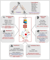
Potential role of necroptosis in human pathological diseases. The necroptosis pathway and its regulatory proteins (RIPK1/-3/MLKL) have been involved in numerous clinical diseases such as cancer, cardiovascular, lung, renal, hepatic, neurodegenerative, and inflammatory diseases. RIPK1, RIPK3, and MLKL-dependent necroptotic cell death have been observed in diseases such as acute kidney and liver injury, autoimmune disease, and cancer. In chronic kidney and lung injury and cardiovascular diseases, RIPK3/MLKL-dependent, but RIPK1-independent, phenotypes have been observed. Furthermore, RIPK1-mediated necroinflammation, independent of necroptosis, has been implicated in neurodegenerative, kidney, renal, sepsis, and autoimmune diseases.
Figure 5
Figure 5
Classification of necroptosis-targeting therapeutic strategies in human clinical diseases.

Similar articles

Cited by

References

    1. Fink S.L., Cookson B.T. Apoptosis, pyroptosis, and necrosis: Mechanistic description of dead and dying eukaryotic cells. Infect. Immun. 2005;73:1907–1916. doi: 10.1128/IAI.73.4.1907-1916.2005. - DOI - PMC - PubMed
    1. Galluzzi L., Vitale I., Abrams J.M., Alnemri E.S., Baehrecke E.H., Blagosklonny M.V., Dawson T.M., Dawson V.L., El-Deiry W.S., Fulda S., et al. Molecular definitions of cell death subroutines: Recommendations of the Nomenclature Committee on Cell Death 2012. Cell Death Differ. 2012;19:107–120. doi: 10.1038/cdd.2011.96. - DOI - PMC - PubMed
    1. Pfeffer C.M., Singh A.T.K. Apoptosis: A Target for Anticancer Therapy. Int. J. Mol. Sci. 2018;19:448. doi: 10.3390/ijms19020448. - DOI - PMC - PubMed
    1. Ge Yan M.E. Thomas Efferth, Multiple cell death modalities and their key features. World Acad. Sci. 2020;2:39–48.
    1. Christofferson D.E., Yuan J. Necroptosis as an alternative form of programmed cell death. Curr. Opin. Cell Biol. 2010;22:263–268. doi: 10.1016/j.ceb.2009.12.003. - DOI - PMC - PubMed

Substances

LinkOut - more resources