. 2022 Oct 22;23(21):12719.
doi: 10.3390/ijms232112719.

Evaluating EcxR for Its Possible Role in Ehrlichia chaffeensis Gene Regulation

Affiliations

Evaluating EcxR for Its Possible Role in Ehrlichia chaffeensis Gene Regulation

Huitao Liu et al. Int J Mol Sci. .

Abstract

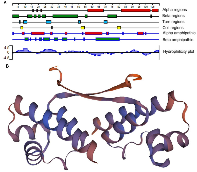
Ehrlichia chaffeensis, a tick-transmitted intraphagosomal bacterium, is the causative agent of human monocytic ehrlichiosis. The pathogen also infects several other vertebrate hosts. E. chaffeensis has a biphasic developmental cycle during its growth in vertebrate monocytes/macrophages and invertebrate tick cells. Host- and vector-specific differences in the gene expression from many genes of E. chaffeensis are well documented. It is unclear how the organism regulates gene expression during its developmental cycle and for its adaptation to vertebrate and tick host cell environments. We previously mapped promoters of several E. chaffeensis genes which are recognized by its only two sigma factors: σ32 and σ70. In the current study, we investigated in assessing five predicted E. chaffeensis transcription regulators; EcxR, CtrA, MerR, HU and Tr1 for their possible roles in regulating the pathogen gene expression. Promoter segments of three genes each transcribed with the RNA polymerase containing σ70 (HU, P28-Omp14 and P28-Omp19) and σ32 (ClpB, DnaK and GroES/L) were evaluated by employing multiple independent molecular methods. We report that EcxR binds to all six promoters tested. Promoter-specific binding of EcxR to several gene promoters results in varying levels of gene expression enhancement. This is the first detailed molecular characterization of transcription regulators where we identified EcxR as a gene regulator having multiple promoter-specific interactions.

Keywords: ehrlichia tick-borne disease; obligate intracellular bacteria; rickettsial gene regulation.


Conflict of interest statement

The authors declare no conflict of interest.

Figures

Figure 1
Figure 1
SouthWestern blotting for the promoters of E. chaffeensis with recombinant DNA-binding proteins. Recombinant DNA-binding proteins were purified and resolved in a 12% SDS-PAGE, transferred to a nitrocellulose membrane and then incubated with 32P-labeled probes to visualize signals by autoradiography. BSA was used as the control non-specific protein control. (A) South-Western blot analysis to assess the interaction of Tr1, EcxR, CtrA, HU and MerR proteins with the full gene promoter segments of p28-Omp19 and p28-Omp14. (B) Full promoters of E. chaffeensis genes, tr1, clpB, groES/L and dnaK, were similarly subjected to the interactions with DNA-binding protein EcxR and Tr1.
Figure 2
Figure 2
E. chaffeensis EcxR secondary structure and tertiary structure predictions. (A) The filled boxes with different colors indicate alpha regions (alpha helical), beta regions (beta strand), turn regions and coli regions, as identified using the Garnier-Osguthorpe-Robson algorithm. (The scale indicates 5-amino-acid intervals.) In the alpha amphipathic and beta amphipathic diagram, the filled boxes indicate regions that are predicted to form alpha helices and beta regions, respectively, and are comprised of amphipathic amino acids, as identified by the Eisenberg algorithm. The Kyte-Doolittle algorithm was used to identify hydrophobic (histogram above the x axis) and hydrophilic (histogram below the axis) regions described at the Hydrophobicity diagram. All of the analyses were carried out using Protean, as a part of the Lasergene software package. (B) Tertiary structure prediction of EcxR. EcxR forming as a homodimer was predicted by using SEWISS-MODEL algorithms.
Figure 3
Figure 3
Electrophoretic Mobility Shift Assays (EMSAs) to assess the interaction of EcxR with different E. chaffeensis gene promoters. (A) From left to right, biotin-labeled probes of clpB, dnaK, groES/L and hup were incubated with recombinant EcxR. High excess cold competitor DNA (unlabeled) was added to show the specificity for protein-promoter interactions. Two DNA segments containing the coding region of dnaK (dnaK-ORF) and ecxR (ecxR-ORF) were used as additional controls to define the specific interactions of EcxR (far right data panels). (B) Biotin-labeled probes of different p28-Omp14 promoter segments (as described in Figure S2A), were used in the EMSA analysis to identify specific regions of these two gene promoters with EcxR. (C) Similarly, biotin-labeled probes of different p28-Omp19 promoter segments were used in the EMSA analysis.
Figure 4
Figure 4
EMSAs to determine binding affinities of two promoter segments. The assays were performed using the biotin-labeled DNA probe of p28-Omp14-P4 (A) or p28-Omp19-P3 (B) incubated with various concentrations of EcxR. DNA probes and EcxR were incubated at room temperature for 20 min before electrophoresis. Samples were run on 5% polyacrylamide-Tris-borate-EDTA (TBE) gel. The amounts of EcxR used in each reaction are indicated at the top of images.
Figure 5
Figure 5
Yeast one-hybrid assay to independently confirm the binding of EcxR to three E. chaffeensis promoters; p28-Omp14 (P14), p28-Omp19 (P19) and tr1 (Ptr1). The β-galactosidase assays were used to quantitate the interaction strength of EcxR binding to three promoters, respectively. Eenzyme activities relaive to the negative control (the empty vector pDEST22) were shown for the three promoters. The values are the means ± standard deviations for three independent biological replicates. Ctrl refers to the negative control (the empty vector pDEST22), while EcxR represents to the expression of AD-EcxR chimeric protein in pDEST22-ecxR. Significant differences from the values for samples lacking the expression of protein EcxR were determined by the t test (* p < 0.05).
Figure 6
Figure 6
Total RNA recovered from synchronously cultured E. chaffeensis in DH82 cells at different time points were used to perform real-time RT-PCR analysis for ecxR gene and normalized against bacterial 16S rRNA. Relative values to the amount at 0 h p.i. are shown. Data indicate mean values ± standard deviations from three independent experiments performed in triplicates. Statistical significance was determined by one-way analysis of variance (ANOVA) followed by Tukey’s multiple-comparison test (* p < 0.05; ** p < 0.01).
Figure 7
Figure 7
Assess the role of EcxR on the transcription of E. chaffeensis promoter-lacZ reporter constructs. β-galactosidase assays were performed to measure the transcriptional activities of lacZ reporter constructs. (A,C) Western blot analyses of samples from β-galactosidase assays were carried out using an anti-His tag antibody to verify the expression of RpoH (σ32) or RpoD (σ70) alone or with EcxR expression. The positions of RpoH, RpoD and EcxR are indicated by arrowheads. (B) The β-galactosidase expression driven by E. chaffeensis promoters constructs encoding clpB, groES/L and dnaK were quantitated in the CAG57101 strain of E. coli expressing E. chaffeensis EcxR and σ32 (EcxR-RpoH) or only E. chaffeensis σ32 (RpoH), which were grown at 30 °C. (D) The β-galactosidase expression driven by E. chaffeensis promoters constructs encoding p28-Omp14, p28-Omp19 and hup were quantitated in the CAG20177 strain of E. coli expressing E. chaffeensis EcxR and σ70 (EcxR-RpoD) or only E. chaffeensis σ70 (RpoD), which were grown at 37 °C. The values are the means ± standard deviations for three independent biological replicates. Significant differences from the values for samples lacking the expression of protein EcxR were determined by the t test (**, p < 0.01; ***, p < 0.001; ns, not significant).
Figure 7
Figure 7
Assess the role of EcxR on the transcription of E. chaffeensis promoter-lacZ reporter constructs. β-galactosidase assays were performed to measure the transcriptional activities of lacZ reporter constructs. (A,C) Western blot analyses of samples from β-galactosidase assays were carried out using an anti-His tag antibody to verify the expression of RpoH (σ32) or RpoD (σ70) alone or with EcxR expression. The positions of RpoH, RpoD and EcxR are indicated by arrowheads. (B) The β-galactosidase expression driven by E. chaffeensis promoters constructs encoding clpB, groES/L and dnaK were quantitated in the CAG57101 strain of E. coli expressing E. chaffeensis EcxR and σ32 (EcxR-RpoH) or only E. chaffeensis σ32 (RpoH), which were grown at 30 °C. (D) The β-galactosidase expression driven by E. chaffeensis promoters constructs encoding p28-Omp14, p28-Omp19 and hup were quantitated in the CAG20177 strain of E. coli expressing E. chaffeensis EcxR and σ70 (EcxR-RpoD) or only E. chaffeensis σ70 (RpoD), which were grown at 37 °C. The values are the means ± standard deviations for three independent biological replicates. Significant differences from the values for samples lacking the expression of protein EcxR were determined by the t test (**, p < 0.01; ***, p < 0.001; ns, not significant).
Figure 8
Figure 8
In vitro transcription analysis of the role of protein EcxR on the transcription of the E. chaffeensis promoters, including clpB, groES/L, dnaK, p28-Omp14 and p28-Omp19. The promoter segments of E. chaffeensis genes, clpB, groES/L, dnaK, p28-Omp14 and p28-Omp19 were cloned upstream to the G-less cassette in pMT504 plasmid vector in the correct orientation and used in the assays with reconstituted RNAP containing E. chaffeensis recombinant σ32 or σ70. In vitro transcription analysis was performed using E. coli RNAP holoenzyme containing E. chaffeensis recombinant σ32 for clpB, groES/L and dnaK, and E. chaffeensis recombinant σ70 for p28-Omp14 and p28-Omp19. Lane 1, template DNA with RNAP holoenzyme with no EcxR added. Lanes 2–3, template DNA with RNAP holoenzyme and with 10 or 20 nM recombinant EcxR, respectively. The abundance of the transcripts for the template in the presence of σ32 or σ70 alone or with recombinant EcxR is captured from the Biotin-14-CTP incorporation in the RNA. The upper panel indicate the transcription products, which were resolved on a 6% denatured polyacrylamide gel containing 7 m urea in 1 × Tris-borate-EDTA buffer. The lower panel indicated the intensity of a band signals in a gel for in vitro transcriptions, as determined using the software ImageJ. The bars show the relative change of transcription products with different concentrations of EcxR as the percentage of transcripts compared to the controls lacking EcxR.

References

    1. Ganguly S., Mukhopadhayay S.K. Tick-borne ehrlichiosis infection in human beings. J. Vector Borne. Dis. 2008;45:273–280. - PubMed
    1. Allan B.F., Goessling L.S., Storch G.A., Thach R.E. Blood meal analysis to identify reservoir hosts for Amblyomma americanum ticks. Emerg. Infect. Dis. 2010;16:433–440. doi: 10.3201/eid1603.090911. - DOI - PMC - PubMed
    1. Dawson J.E., Biggie K.L., Warner C.K., Cookson K., Jenkins S., Levine J.F., Olson J.G. Polymerase chain reaction evidence of Ehrlichia chaffeensis, an etiologic agent of human monocytic ehrlichiosis, in dogs, from southeast Virginia. Am. J. Vet. Res. 1996;57:1175–1179. - PubMed
    1. Lockhart J.M., Davidson W.R., Stallknecht D.E., Dawson J.E., Howerth E.W. Isolation of Ehrlichia chaffeensis from wild white-tailed deer (Odocoileus virginianus) confirms their role as natural reservoir hosts. J. Clin. Microbiol. 1997;35:1681–1686. doi: 10.1128/jcm.35.7.1681-1686.1997. - DOI - PMC - PubMed
    1. Breitschwerdt E.B., Hegarty B.C., Hancock S.I. Sequential evaluation of dogs naturally infected with Ehrlichia canis, Ehrlichia chaffeensis, Ehrlichia equi, Ehrlichia ewingii, or Bartonella vinsonii. J. Clin. Microbiol. 1998;36:2645–2651. doi: 10.1128/JCM.36.9.2645-2651.1998. - DOI - PMC - PubMed

Substances

LinkOut - more resources