. 2022 Oct 23;23(21):12764.
doi: 10.3390/ijms232112764.

Molecular Mechanism of Sirtuin 1 Modulation by the AROS Protein

Affiliations

Molecular Mechanism of Sirtuin 1 Modulation by the AROS Protein

Sandra Weiss et al. Int J Mol Sci. .

Abstract

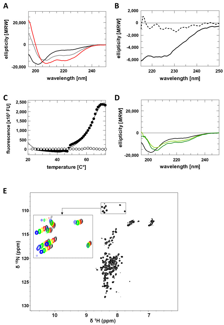
The protein lysine deacylases of the NAD+-dependent Sirtuin family contribute to metabolic regulation, stress responses, and aging processes, and the human Sirtuin isoforms, Sirt1-7, are considered drug targets for aging-related diseases. The nuclear isoform Sirt1 deacetylates histones and transcription factors to regulate, e.g., metabolic adaptations and circadian mechanisms, and it is used as a therapeutic target for Huntington's disease and psoriasis. Sirt1 is regulated through a multitude of mechanisms, including the interaction with regulatory proteins such as the inhibitors Tat and Dbc1 or the activator AROS. Here, we describe a molecular characterization of AROS and how it regulates Sirt1. We find that AROS is a partly intrinsically disordered protein (IDP) that inhibits rather than activates Sirt1. A biochemical characterization of the interaction including binding and stability assays, NMR spectroscopy, mass spectrometry, and a crystal structure of Sirtuin/AROS peptide complex reveal that AROS acts as a competitive inhibitor, through binding to the Sirt1 substrate peptide site. Our results provide molecular insights in the physiological regulation of Sirt1 by a regulator protein and suggest the peptide site as an opportunity for Sirt1-targeted drug development.

Keywords: Sirt1; activator; deacetylase; inhibitor; regulation.


Conflict of interest statement

The authors declare no conflict of interest.

Figures

Figure 1
Figure 1
Sequence analysis, recombinant preparation and folding analysis of AROS protein. (A) Prediction of AROS secondary structure elements with PSIPRED (H/pink: α-helix; C/grey: coil). Confidence of prediction: blue (high), white (low). (B) Disorder prediction for human AROS using DISOPRED (black) and PONDR (red). (C) SEC elution profile of purified, refolded AROS (red) and of globular standard proteins (black; MW indicated). Inset: comparison of AROS elution profiles without additive (red) and with 4 M urea (dotted line) or 4 M GdnHCl (dashed line). MW values from comparison to standard proteins are indicated. (D) Blue native PAGE of refolded, purified AROS.
Figure 2
Figure 2
Structural characterization of AROS. (A) CD spectra of AROS in absence and presence of TFE (0% black, 10% grey, 20% red). (B) CD spectra of AROS in absence (solid line) and presence of 6 M urea (dashed line). (C) Thermal denaturation of AROS in absence (white) and presence of 50% TFE (black). (D) CD spectra of AROS in absence and presence of glycerol (0% black, 40% light green, 50% dark green). (E) [1H,15N]-HSQC spectra of AROS with an enlarged view of glycine signals recorded at different temperatures (25 °C black, 20 °C red, 15 °C yellow, 10 °C green, 5 °C blue).
Figure 3
Figure 3
Sirt1 inhibition by AROS. (A) Thermal denaturation of the Sirt1/AROS complex in 20 mM Tris/HCl pH 7.0; 120 mM NaCl; 0.08% Tween 20 (white), or in 50 mM Tris/HCl pH 7.5 (grey), or in 50 mM Na2HPO4/NaH2PO4 pH 7.0 (black). (B) Dose-dependent inhibition of fl-Sirt1 (black) and mini-Sirt1 (white) by AROS in the FdL deacetylation assay. (n = 3; error bars: SD) (C) Substrate peptide titrations of Sirt1 in FdL activity assays in presence (white) and absence (black) of 40 µM AROS. (n = 3; error bars: SD) (D) NAD+ titrations of Sirt1 in FdL activity assays in presence (white) and absence (black) of 40 µM AROS. (n = 3; error bars: SD).
Figure 4
Figure 4
Mapping of the Sirt1/AROS interaction interfaces. (A) 2D [1H,15N]-HSQC spectra of the titration of 150 µM 15N-AROS with mini-Sirt1. Spectra corresponding to molar ratios 1:0, 1:0.5, and 1:1 are in black, cyan, and red, respectively. Examples of changed and unchanged signals are highlighted, and assigned signals are labeled (see Supplementary Figure S3 for mapping of changed signals on the sequence). (B) Crosslinking experiments with DSSO showing complex formation of Sirt1 with full-length AROS. (C) Effects of AROS on the deacetylase activities of mini-Sirt1 (black), Sirt2 (grey), and Sirt3 (white) and the desuccinylase activity of Sirt5 (striped). (n = 3; error bars: SD) (D,E) Crosslinking experiments with DSSO showing complex formation of AROS (D) with Sirt3 and (E) with Sirt5. (F) Location of Sirt5 residues identified as cross-linking sites indicated as sticks and labeled. The figure was generated from the crystal structure of a Sirt5 complex with substrate peptide and NAD+ analog (PDB ID 4G1C) [28].
Figure 5
Figure 5
Crystal structure of a Sirt3/AROS peptide complex and modeling of the Sirt1/AROS complex. (A) 2D-NMR titration of 15N-labeled AROS with Sirt3, analogous to the experiment with Sirt1 (see Figure 4A). Shown are molar ratios 1:0 and 1:1 in black and red, respectively. (B) Overall structure of the complex between Sirt3 and AROS peptide 62-71 (green sticks). 2Fo-Fc electron density (blue) for AROS62-71 is contoured at 1σ. (C) Overlay of Sirt3 (gray cartoon) in complex with AROS62-71 (green sticks) and miniSirt1 (wheat cartoon) in complex with an acetylated p53 peptide (magenta sticks), generated through secondary structure matching for the Sirtuin cores (other overlaid Sirtuin/peptide complexes omitted for clarity). Enlarged view shows interactions of AROS62-71 and p53 with the Sirtuin active site. The Sirt3 complex is shifted slightly up compared to the Sirt1 complex for visibility. (D) Structure prediction for AROS using the program RaptorX (wheat cartoon). The original orientation of the C-terminus was changed manually (arrow) to obtain a more extended conformation (green cartoon) for docking onto Sirt1. (E) Modeling of the Sirt1 (cyan; pdb entry: 4zzh) interaction with AROS (green) based on the model in panel D, the Sirt3/AROS crystal structure, and our cross-linking results. Residues implicated in the interaction through cross-links (red) and the catalytic Sirt1 His363 (yellow) are shown as sticks, and regions implicated in the interaction through NMR are circled (purple).

References

    1. Rauh D., Fischer F., Gertz M., Lakshminarasimhan M., Bergbrede T., Aladini F., Kambach C., Becker C.F.W., Zerweck J., Schutkowski M., et al. An acetylome peptide microarray reveals specificities and deacetylation substrates for all human sirtuin isoforms. Nat. Commun. 2013;4:2327. - PubMed
    1. Vassilopoulos A., Fritz K.S., Petersen D.R., Gius D. The human sirtuin family: Evolutionary divergences and functions. Hum. Genom. 2011;5:1–12. - PMC - PubMed
    1. Morris B.J. Seven sirtuins for seven deadly diseases of aging. Free Radic. Biol. Med. 2013;56:133–171. doi: 10.1016/j.freeradbiomed.2012.10.525. - DOI - PubMed
    1. Donmez G., Outeiro T.F. SIRT1 and SIRT2: Emerging targets in neurodegeneration. EMBO Mol. Med. 2013;5:344–352. doi: 10.1002/emmm.201302451. - DOI - PMC - PubMed
    1. Dai H., Sinclair D.A., Ellis J.L., Steegborn C. Sirtuin activators and inhibitors: Promises, achievements, and challenges. Pharmacol. Ther. 2018;188:140–154. doi: 10.1016/j.pharmthera.2018.03.004. - DOI - PMC - PubMed

LinkOut - more resources