Review
. 2022 Oct 25;23(21):12841.
doi: 10.3390/ijms232112841.

Phosphorylated Tau in Alzheimer's Disease and Other Tauopathies

Affiliations
Review

Phosphorylated Tau in Alzheimer's Disease and Other Tauopathies

Priyanka Rawat et al. Int J Mol Sci. .

Abstract

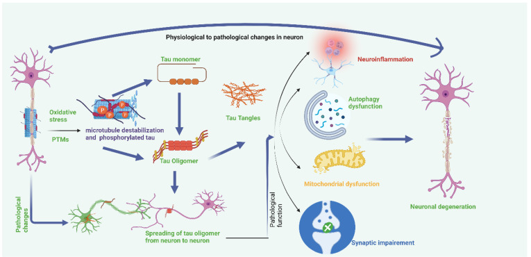
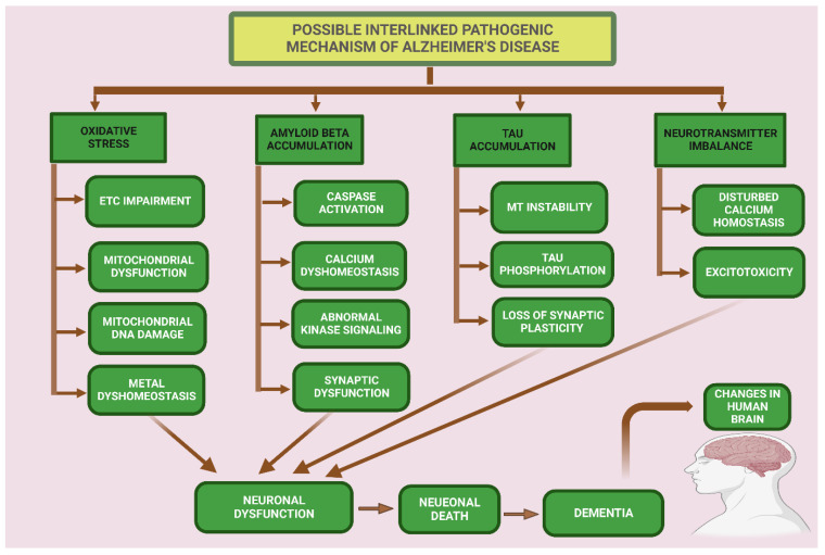
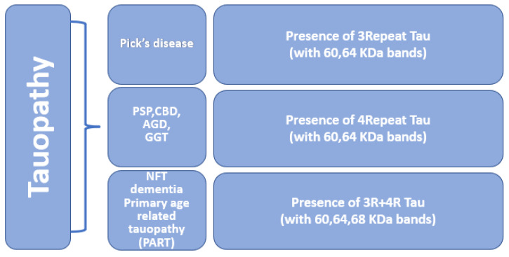
Alzheimer's disease (AD) is the leading cause of dementia in elderly people. Amyloid beta (Aβ) deposits and neurofibrillary tangles are the major pathological features in an Alzheimer's brain. These proteins are highly expressed in nerve cells and found in most tissues. Tau primarily provides stabilization to microtubules in the part of axons and dendrites. However, tau in a pathological state becomes hyperphosphorylated, causing tau dysfunction and leading to synaptic impairment and degeneration of neurons. This article presents a summary of the role of tau, phosphorylated tau (p-tau) in AD, and other tauopathies. Tauopathies, including Pick's disease, frontotemporal dementia, corticobasal degeneration, Alzheimer's disease, argyrophilic grain disease, progressive supranuclear palsy, and Huntington's disease, are the result of misprocessing and accumulation of tau within the neuronal and glial cells. This article also focuses on current research on the post-translational modifications and genetics of tau, tau pathology, the role of tau in tauopathies and the development of new drugs targeting p-tau, and the therapeutics for treating and possibly preventing tauopathies.

Keywords: Alzheimer’s disease; amyloid beta; dementia; oxidative stress; phosphorylated tau; tauopathy.


Conflict of interest statement

The authors declare no conflict of interest.

Figures

Figure 1
Figure 1
Non-modifiable and modifiable risk factors for Alzheimer’s disease. Non-modifiable factors that may cause AD are age, sex, and defective genetics. Modifiable risk factors (e.g., obesity, smoking, drinking, heart disease, physical inactivity) can play a major role as preventive factors.
Figure 2
Figure 2
MAPT gene and 6-isoforms structure: The tau gene is located on chromosome 17q21 and contains 16 exons. Six isoforms of tau are generated by alternate splicing of the exons 2, 3, and 10 of the MAPT gene. The N terminal domain, N1 and N2, are produced from exons 2 and 3. These isoforms differ in length from 352–441 amino acids.
Figure 3
Figure 3
Oxidative stress and post-translational modifications cause tau hyperphosphorylation, which causes instability in microtubules. Microtubule instability causes the formation of tau tangles from the tau monomer and tau oligomer. Accumulation of tau tangles causes neuroinflammation, autophagy dysfunction, mitochondrial dysfunction, and synaptic impairment, which results in neuronal damage. Tau oligomers also spread from neuron to neuron.
Figure 4
Figure 4
Pathogenic hypothesis for neuronal dysfunction in Alzheimer’s disease. Mitochondrial oxidative stress condition accelerates the electron transport system to generate more reactive oxidative stress and cause oxidative damage in brain cells. Aβ accumulation causes caspase activation of abnormal kinase signaling and synaptic impairment. Caspase activation increases tau phosphorylation. Tau accumulation in cells causes microtubule instability which causes tau phosphorylation. A neurotransmitter imbalance causes disturbed calcium homeostasis and excitotoxicity. Oxidative stress, Aβ accumulation, tau accumulation, and neurotransmitter imbalances result in neuronal dysfunction, which causes neuronal death and leads to dementia.
Figure 5
Figure 5
Molecular classification of primary tauopathy. Tauopathies are divided into three categories based on the presence of repeat isoforms. 3-repeat tauopathy is linked with Pick’s disease. The presence of 4-repeat isoforms is linked with PSP, CBD, AGD, and GGT. The presence of both 3-repeat and 4-repeat tau isoforms is associated with primary age-related tauopathy and tangles-only dementia.

References

    1. John A., Reddy P.H. Synaptic basis of Alzheimer’s disease: Focus on synaptic amyloid beta, P-tau and mitochondria. Ageing Res. Rev. 2021;65:101208. doi: 10.1016/j.arr.2020.101208. - DOI - PMC - PubMed
    1. Morton H., Kshirsagar S., Orlov E., Bunquin L.E., Sawant N., Boleng L., George M., Basu T., Ramasubramanian B., Pradeepkiran J.A. Defective mitophagy and synaptic degeneration in Alzheimer’s disease: Focus on aging, mitochondria and synapse. Free Radic. Biol. Med. 2021;172:652–667. doi: 10.1016/j.freeradbiomed.2021.07.013. - DOI - PubMed
    1. Selkoe D.J. Alzheimer’s disease: Genes, proteins, and therapy. Physiol. Rev. 2001;81:741–766. doi: 10.1152/physrev.2001.81.2.741. - DOI - PubMed
    1. Sinsky J., Pichlerova K., Hanes J. Tau protein interaction partners and their roles in Alzheimer’s disease and other tauopathies. Int. J. Mol. Sci. 2021;22:9207. doi: 10.3390/ijms22179207. - DOI - PMC - PubMed
    1. Goedert M., Jakes R. Mutations causing neurodegenerative tauopathies. Biochim. Et Biophys. Acta (BBA)-Mol. Basis Dis. 2005;1739:240–250. doi: 10.1016/j.bbadis.2004.08.007. - DOI - PubMed