Review
. 2022 Oct 26;23(21):12906.
doi: 10.3390/ijms232112906.

The Role of Inflammation in Cardiovascular Disease

Affiliations
Review

The Role of Inflammation in Cardiovascular Disease

Michael Y Henein et al. Int J Mol Sci. .

Abstract

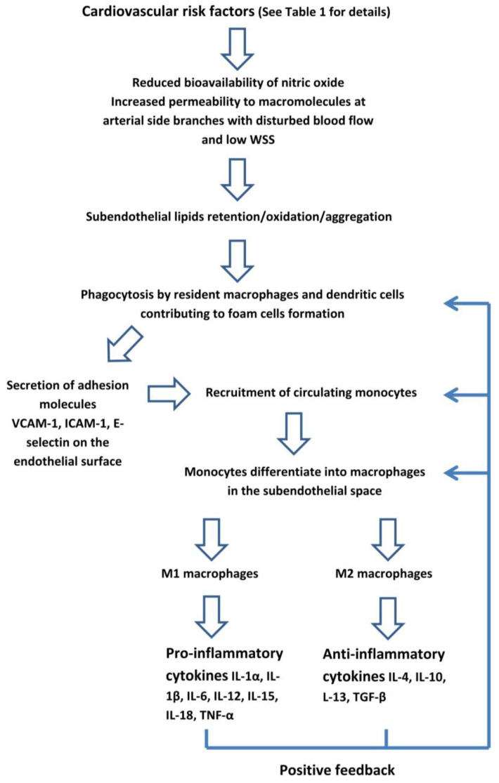
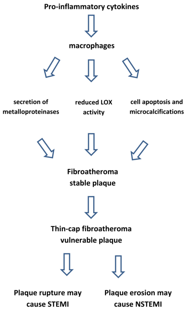
Atherosclerosis is a chronic inflammatory disease, in which the immune system has a prominent role in its development and progression. Inflammation-induced endothelial dysfunction results in an increased permeability to lipoproteins and their subendothelial accumulation, leukocyte recruitment, and platelets activation. Recruited monocytes differentiate into macrophages which develop pro- or anti-inflammatory properties according to their microenvironment. Atheroma progression or healing is determined by the balance between these functional phenotypes. Macrophages and smooth muscle cells secrete inflammatory cytokines including interleukins IL-1β, IL-12, and IL-6. Within the arterial wall, low-density lipoprotein cholesterol undergoes an oxidation. Additionally, triglyceride-rich lipoproteins and remnant lipoproteins exert pro-inflammatory effects. Macrophages catabolize the oxidized lipoproteins and coalesce into a lipid-rich necrotic core, encapsulated by a collagen fibrous cap, leading to the formation of fibro-atheroma. In the conditions of chronic inflammation, macrophages exert a catabolic effect on the fibrous cap, resulting in a thin-cap fibro-atheroma which makes the plaque vulnerable. However, their morphology may change over time, shifting from high-risk lesions to more stable calcified plaques. In addition to conventional cardiovascular risk factors, an exposure to acute and chronic psychological stress may increase the risk of cardiovascular disease through inflammation mediated by an increased sympathetic output which results in the release of inflammatory cytokines. Inflammation is also the link between ageing and cardiovascular disease through increased clones of leukocytes in peripheral blood. Anti-inflammatory interventions specifically blocking the cytokine pathways reduce the risk of myocardial infarction and stroke, although they increase the risk of infections.

Keywords: atherosclerosis; cerebral artery aneurysm; coronary artery disease; coronary atherosclerotic plaque; inflammation; stroke.


Conflict of interest statement

The authors declare no conflict of interest.

Figures

Figure 1
Figure 1
The early phase of atherosclerotic lesions starts with endothelial dysfunction which triggers a low-grade inflammatory response. WSS: wall shear stress. VCAM: vascular cell adhesion molecule. ICAM: intercellular adhesion molecule.
Figure 2
Figure 2
Selective angiography of the left coronary artery. Severe focal atherosclerotic narrowing of the proximal segment of left anterior descending coronary artery.
Figure 3
Figure 3
Artery-to-artery embolism and in situ thrombotic occlusion of the middle cerebral artery due to intracranial atherosclerotic disease. Hypertensive 67-year-old man. Diffusion-weighted magnetic resonance imaging demonstrates multiple cortical-subcortical ischemic lesions in the territory of the right middle cerebral artery (AC). Digital subtraction angiography shows an occlusion of the M1 segment of the right middle cerebral artery (D) and its complete recanalization after mechanical thrombectomy (E). The magnified oblique projection after the recanalization (F) reveals an underlying atherosclerotic plaque at the site of the previous occlusion (arrow) and additional stenotic lesions along the course of the right anterior cerebral artery (arrowhead).
Figure 4
Figure 4
Internal carotid artery occlusion, digital subtraction angiography: acute thrombotic occlusion of the left internal carotid artery (A) causing sudden neurologic deficit in a 77-year-old patient. The serigraphy performed after mechanical thrombectomy and recanalization of the artery (B) highlights an ulcerated atherosclerotic plaque of the carotid bulb (arrow). After the administration of intravenous boluses of antiplatelet agents and heparin, a self-expanding stent (arrow) was placed in correspondence to the ulcerated plaque (C).
Figure 5
Figure 5
A 61 year old man referred for Non-ST-segment elevation myocardial infarction (NSTEMI). Coronary angiography showed no significant coronary lesions. Left circumflex showed haziness at the proximal segment (panel (A), white arrow). The OCT pull-back showed a fractured plaque (panel (B), 1, white arrow) associated with a lipid pool (panel (B), 1 and 2, “L”); thin-cap fibro-atheroma and active macrophages are easily detected because of their typical bright line (2, white arrows) or spot images, within a fibro-lipidic plaque (3, white arrows).
Figure 6
Figure 6
A 54 year old woman after a scheduled angiogram and OCT pull-back 3 years after an acute coronary syndrome. During index procedure 2 bioresorbable vascular scaffolds (BVS) were implanted. Coronary angiography showed satisfactory angiographical result (panel (A), white line). OCT pull-back confirmed a complete struts coverage and an acceptable lumen area (panel (B)), even at the overlapping site (2). Black boxes are typical OCT images of BVS (1,2,3 and 4). Some black box inclusions are detected (1,2 and 3 – white arrows) representing scaffold reabsorption processes. Some areas immediately below the boxes showed bright spots indicating inflammation with macrophages activation (1 and 2 - underlined area). These bright spots (3 and 4, white triangle) have been detected closely to calcium arch (3, stars) and in a fibro-lipidic plaque (4).
Figure 7
Figure 7
Polyvascular disease: right frontal lobar haemorrhage due to cerebral amyloid angiopathy (A) in an 82-year-old man with a prosthetic aortic valve and extensive calcifications of the aortic arch and along the descending thoracic aorta (B). Three-dimensional digital subtraction angiography reconstructions of the same patient (C,D) show diffuse arterial dysplasia with ecstatic origin of an inferior temporal branch (arrow), a dysmorphic aneurysm of the Sylvian bifurcation of the middle cerebral artery (arrowhead), and an infra-millimetric aneurysm of the anterior communicating artery (*).
Figure 8
Figure 8
Inflammation makes atherosclerotic plaque vulnerable. LOX: lysyl oxidase enzyme. STEMI: ST-segment elevation myocardial infarction. NSTEMI: Non-ST-segment elevation myocardial infarction.

References

    1. Libby P. The changing landscape of atherosclerosis. Nature. 2021;592:524–533. doi: 10.1038/s41586-021-03392-8. - DOI - PubMed
    1. Libby P., Hansson G.K. From Focal Lipid Storage to Systemic Inflammation: JACC Review Topic of the Week. J. Am. Coll. Cardiol. 2019;74:1594–1607. doi: 10.1016/j.jacc.2019.07.061. - DOI - PMC - PubMed
    1. Hansson G.K. Inflammation, Atherosclerosis, and Coronary Artery Disease. N. Engl. J. Med. 2005;352:1685–1695. doi: 10.1056/NEJMra043430. - DOI - PubMed
    1. Ruparelia N., Choudhury R. Inflammation and atherosclerosis: What is on the horizon? Heart. 2020;106:80–85. doi: 10.1136/heartjnl-2018-314230. - DOI - PubMed
    1. Libby P. Inflammation during the life cycle of the atherosclerotic plaque. Cardiovasc. Res. 2021;117:2525–2536. doi: 10.1093/cvr/cvab303. - DOI - PMC - PubMed

MeSH terms