. 2022 Oct 28;23(21):13130.
doi: 10.3390/ijms232113130.

Benzoylpaeoniflorin Activates Anti-Inflammatory Mechanisms to Mitigate Sepsis in Cell-Culture and Mouse Sepsis Models

Affiliations

Benzoylpaeoniflorin Activates Anti-Inflammatory Mechanisms to Mitigate Sepsis in Cell-Culture and Mouse Sepsis Models

Chaeyeong Kim et al. Int J Mol Sci. .

Abstract

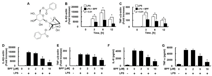
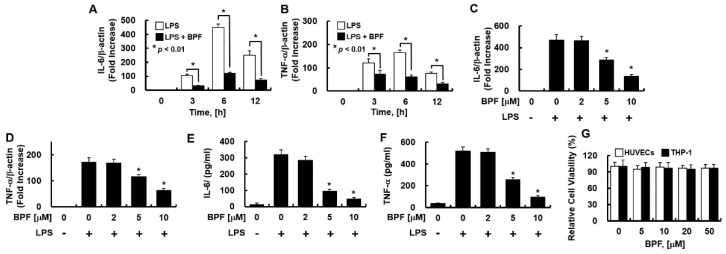
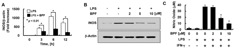
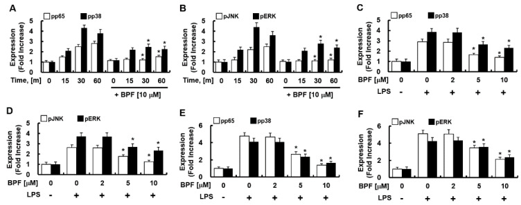
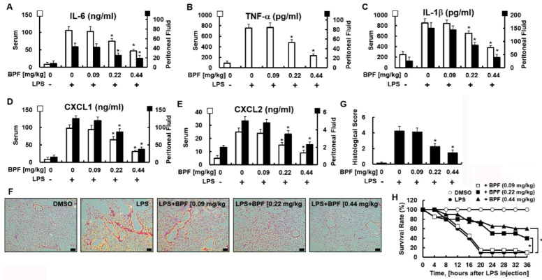
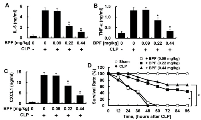
Xuebijing injection (XBJI) (comprising of five herbs) is a widely used traditional Chinese medicine for sepsis treatment. However, the bioactive components of XBJI and the mechanisms responsible for its sepsis-mitigating action have not been experimentally determined. One of the main bioactive compounds in XBJI-benzoylpaeoniflorin (BPF)-inhibits the expressions of key mediators of inflammation such as nuclear factor kappa B (NF-κB), cyclooxygenase-1 (COX-1), and COX-2. However, its effects on sepsis have not been determined yet. Therefore, here, we investigated the immunomodulatory effect of BPF on severely inflamed endothelial cells, THP-1 macrophages, peritoneal macrophages, and mice. Human umbilical vein endothelial cells (HUVECs) and THP-1-macrophages were activated using lipopolysaccharide (LPS) after pretreatment with BPF. Subsequently, changes in the expression profiles of pro-inflammatory molecules including inducible nitric oxide synthase (iNOS), tumor necrosis factor (TNF)-α, and interleukin (IL)-6 were determined using quantitative real-time polymerase chain reaction (qPCR) and Western blot analysis. Furthermore, we monitored the phosphorylation of NF-kB and mitogen-activated protein kinases (MAPKs) to determine their activation levels. Using the LPS-induced mouse model of sepsis, we studied the effects of BPF on inflammatory cytokine production, pulmonary histopathology, and survival rates. Finally, we evaluated whether BPF protects against cecal ligation and puncture (CLP)-induced sepsis, as it closely mimics human sepsis. BPF pretreatment inhibited LPS-induced increase in mRNA and protein levels of iNOS, TNF-α, and IL-6 in HUVECs and THP-1-macrophages. It also suppressed LPS-mediated phosphorylation of p65, p38, JNK, and ERK. Mice with LPS-induced-sepsis who were treated with BPF had lower serum levels of IL-6, TNF-α, IL-1β, CXCL1, and CXCL2 than the control mice treated with BPF. Histopathology revealed that BPF treatment alleviated LPS-induced lung damage. In addition, in mice given a lethal dose of LPS, BPF treatment showed a dose-dependent improvement in survival rates. BPF treatment dose-dependently inhibited the LPS-induced IL-6, TNF-α, and CXCL1 production in peritoneal macrophages. BPF treatment also dose-dependently improved the survival rates in mice with CLP-induced sepsis. These results show that BPF alleviates LPS-stimulated septic conditions and protects mice from CLP-induced sepsis. Our research marks BPF as a potential drug in the treatment of sepsis and various inflammatory diseases.

Keywords: CLP; benzoylpaeoniflorin; endothelium; inflammation; lipopolysaccharide; sepsis.


Conflict of interest statement

The authors declare no conflict of interest.

Figures

Figure 1
Figure 1
Benzoylpaeoniflorin (BPF) inhibits the expression of inflammatory cytokines in LPS-stimulated HUVECs. (A) Structure of BPF; (B,C) HUVECs were pretreated with BPF (10 μM for 6 h), then LPS (100 ng/mL) was added. After the indicated times, total mRNA was extracted and the expression level of each cytokine and β-actin were determined using real-time PCR; (D,E) HUVECs were pretreated with the indicated concentrations of BPF for 6 h, then LPS (100 ng/mL) was added. After 12 h, total mRNA was extracted and the expression level of each cytokine and β-actin was determined using real-time PCR; (F,G) HUVECs were pretreated with BPF at the indicated concentrations for 6 h, then LPS (100 ng/mL) was added. After 24 h, the cell culture supernatant was harvested, and cytokine levels were determined by ELISA. All results are shown as means ± SD of three different experiments with triple samples. * p < 0.5 compared to LPS only (DG).
Figure 2
Figure 2
BPF inhibits the expression of inflammatory cytokines in LPS-stimulated THP-1-macrophages. (A,B) THP-1-macrophages were pretreated with BPF (10 μM, for 6 h), then LPS (100 ng/mL) was added. After the indicated times, total mRNA was extracted and the expression level of each cytokine and β-actin were determined using real-time PCR; (C,D) THP-1-macrophages were pretreated with the indicated concentrations of BPF for 6 h, then LPS (100 ng/mL) was added. After 12 h, the total mRNA was extracted and the expression level of each cytokine and β-actin was determined using real-time PCR; (E,F) THP-1-macrophages were pretreated with BPF at the indicated concentrations for 6 h, then LPS (100 ng/mL) was added. After 24 h, the cell culture supernatant was harvested, and cytokine levels were determined using ELISA. (G) HUVECs or THP-1-macrophages were treated with the indicated concentrations of BPF for 48 h. Cell viability was determined using the MTT assay. All results are shown as means ± SD of three different experiments with triple samples. * p < 0.5 compared to LPS only (DF).
Figure 3
Figure 3
BPF inhibits the expression of iNOS and the production of nitric oxide in HUVECs. (A) HUVECs were pretreated with BPF (10 μM for 6 h), then LPS (100 ng/mL) was added. After the indicated times, total mRNA was extracted and the expression level of iNOS and β-actin was determined using real-time PCR; (B) HUVECs were pretreated with the indicated concentrations of BPF for 6 h, then LPS (100 ng/mL) was added. After 24 h, the cellular protein was extracted and iNOS and β-actin levels were determined using Western blots; (C) HUVECs were pretreated with the indicated concentrations of BPF for 6 h, then LPS (100 ng/mL) and IFN-γ (200 ng/mL) were added under the indicated conditions. After 24 h, the cell culture supernatant was harvested, and nitric oxide levels were determined using the Griess reaction assay (A,C). All results are shown as means ± SD of three different experiments with triple samples. * p < 0.5 compared to LPS + IFN-γ (C); (B) The results are from one representative experiment of three independent experiments.
Figure 4
Figure 4
BPF inhibits the phosphorylation of NF-κB and MAP kinase in LPS-stimulated macrophages. (A,B) HUVECs were pretreated with BPF (10 μM for 6 h), then LPS (100 ng/mL) was added. Cellular protein was extracted at the indicated times; (C,D) HUVECs were pretreated with the indicated concentrations of BPF for 6 h, then LPS (100 ng/mL) was added. After 1 h, the cellular protein was extracted. (E,F) THP-1-macrophages cells were pretreated with the indicated concentrations of BPF for 6 h, then LPS (100 ng/mL) was added. After 1 h, the cellular protein was extracted (AC) The levels of the indicated proteins were determined using ELISA. All results are shown as the means ± SD of three different experiments with triple samples. * p < 0.5 compared to LPS only.
Figure 5
Figure 5
BPF alleviates excessive cytokine production and tissue damage of the lungs and improves the survival rate in the LPS-induced mouse septic shock model. (AE) Mice were injected with 2.5 mg/kg of LPS (intraperitoneal injection) and DMSO or BPF (0.09, 0.22 or 0.44 mg/kg, intravenous injection). After 4 h, the mice were euthanized. Blood and peritoneal fluid were collected and the levels of the indicated inflammatory cytokines were determined using ELISA (n = 5). (F,G) Mice were injected with 2.5 mg/kg LPS (intraperitoneal injection) and DMSO or BPF (0.09, 0.22 or 0.44 mg/kg, intravenous injection). After 24 h, the mice were euthanized and the left lung lobes were collected and fixed in formalin, followed by staining with H&E. H&E staining of lung tissues from each group was conducted, and representative images from three independent experiments conducted on three different days are shown. The bar represents 200 μm. Histopathological scores were obtained using an arbitrary scoring index based on the degree of inflammatory cell infiltration and the extent of the lesion area (n = 5). (H) Mice were injected with 25 mg/kg of LPS (intraperitoneal injection) and DMSO or BPF (0.09, 0.22 or 0.44 mg/kg, intravenous injection). The survival of the mice was monitored every 4 h for 36 h and the survival rates were expressed as a percentage (n = 20). All results are shown as the means ± SD of three different experiments with triple samples. * p < 0.5 compared to LPS only.
Figure 6
Figure 6
BPF suppresses CLP-induced cytokine production of peritoneal macrophages and improves the mortality of CLP mice. (AC) CLP-operated mice were injected with DMSO or BPF (0.09, 0.22, or 0.44 mg/kg, intravenous injection). After 12 h, the mice were euthanized. Peritoneal macrophages were collected from CLP-operated mice and the levels of the indicated inflammatory cytokines were determined using ELISA (n = 5). All results are shown as means ± SD of three different experiments with triple samples. * p < 0.1 compared to CLP only; (D) CLP surgery was conducted on mice and the indicated reagents were administered every 24 h (n = 20). The survival of mice was monitored for 96 h. * p < 0.5.

References

    1. Singer M., Deutschman C.S., Seymour C.W., Shankar-Hari M., Annane D., Bauer M., Bellomo R., Bernard G.R., Chiche J.D., Coopersmith C.M. The Third International Consensus Definitions for Sepsis and Septic Shock (Sepsis-3) JAMA. 2016;315:801–810. doi: 10.1001/jama.2016.0287. - DOI - PMC - PubMed
    1. Dickson K., Lehmann C. Inflammatory Response to Different Toxins in Experimental Sepsis Models. Int. J. Mol. Sci. 2019;20:4341. doi: 10.3390/ijms20184341. - DOI - PMC - PubMed
    1. Ziesmann M.T., Marshall J.C. Multiple Organ Dysfunction: The Defining Syndrome of Sepsis. Surg. Infect. Larchmt. 2018;19:184–190. doi: 10.1089/sur.2017.298. - DOI - PubMed
    1. Bae J.S. Role of high mobility group box 1 in inflammatory disease: Focus on sepsis. Arch. Pharm. Res. 2012;35:1511–1523. doi: 10.1007/s12272-012-0901-5. - DOI - PubMed
    1. Nathan C. Points of control in inflammation. Nature. 2002;420:846–852. doi: 10.1038/nature01320. - DOI - PubMed