. 2022 Oct 30;23(21):13224.
doi: 10.3390/ijms232113224.

Blockage of KHSRP-NLRP3 by MCC950 Can Reverse the Effect of Manganese-Induced Neuroinflammation in N2a Cells and Rat Brain

Affiliations

Blockage of KHSRP-NLRP3 by MCC950 Can Reverse the Effect of Manganese-Induced Neuroinflammation in N2a Cells and Rat Brain

Sharad Singh et al. Int J Mol Sci. .

Abstract

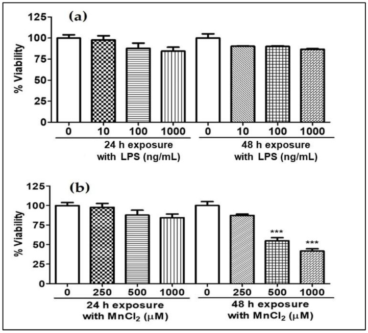
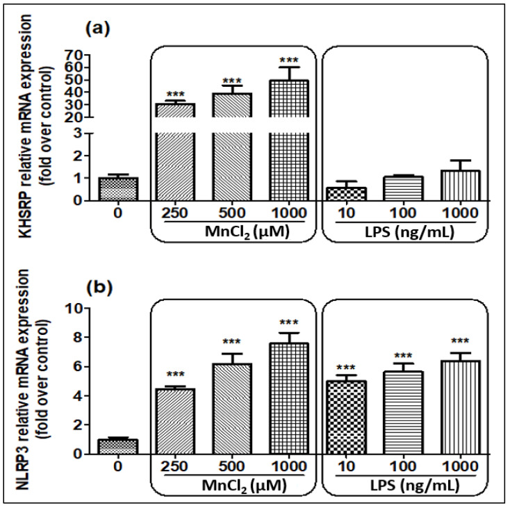
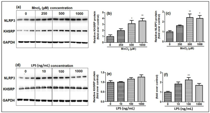
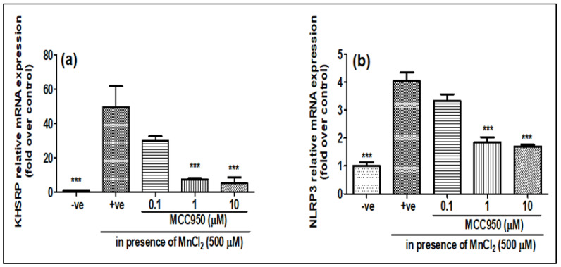
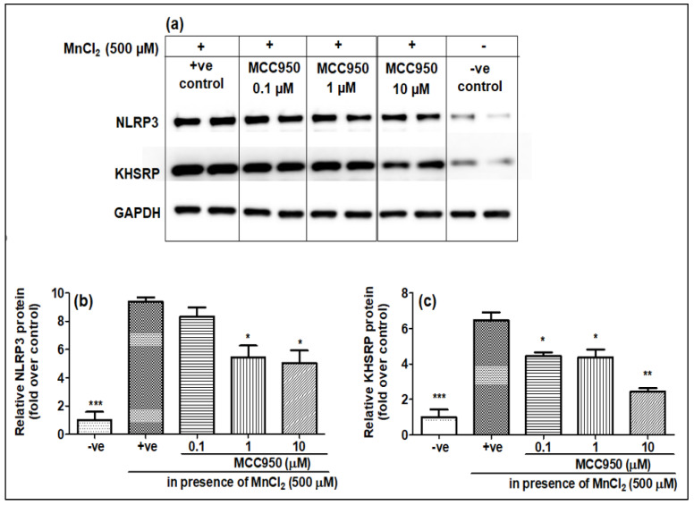
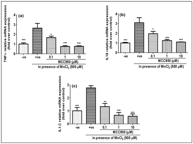
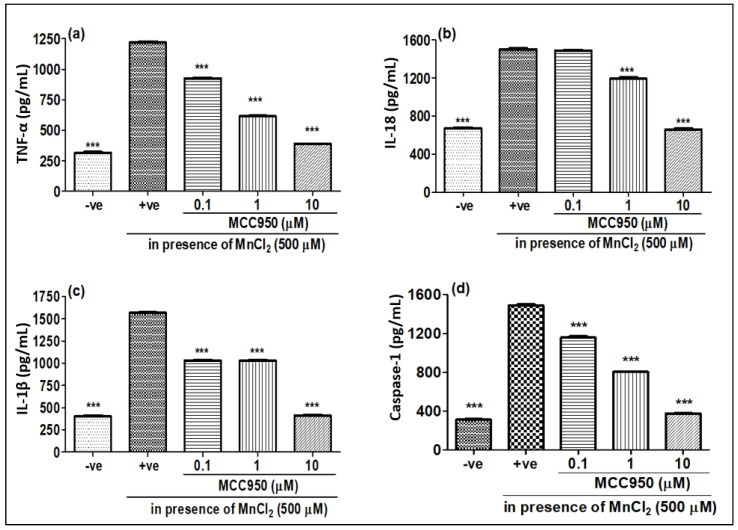
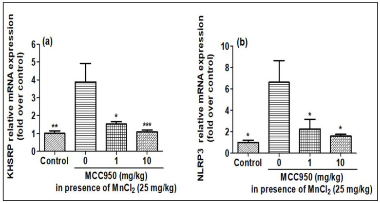
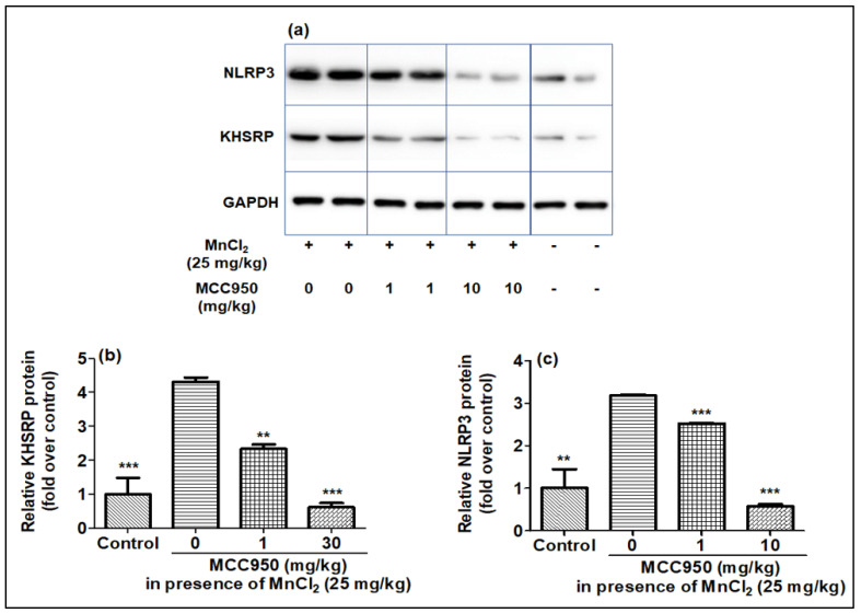
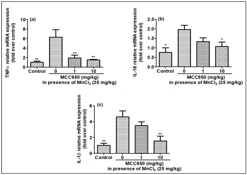
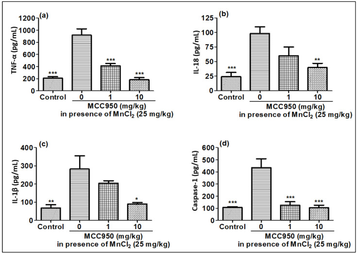
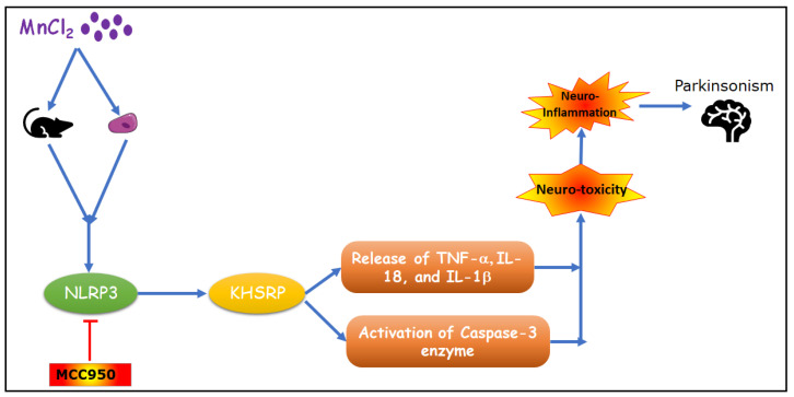
Manganese neurotoxicity has been reported to cause a neurodegenerative disease known as parkinsonism. Previous reports have shown that the expression of the KH-type splicing regulatory protein (KHSRP), a nucleic acid-binding protein, and NLRP3 is increased upon Mn exposure. However, the relation between these two during Mn toxicity has not been fully deduced. The mouse neuroblastoma (N2a) and SD rats are treated with LPS and MnCl2 to evaluate the expression of KHSRP and NLRP3. Further, the effect of the NLRP3 inhibitor MCC950 is checked on the expression of NLRP3, KHSRP and pro-inflammatory markers (TNFα, IL-18 and IL-1β) as well as the caspase-1 enzyme. Our results demonstrated an increment in NLRP3 and KHSRP expression post-MnCl2 exposure in N2a cells and rat brain, while on the other hand with LPS exposure only NLRP3 expression levels were elevated and KHSRP was found to be unaffected. An increased expression of KHSRP, NLRP3, pro-inflammatory markers and the caspase-1 enzyme was observed to be inhibited with MCC950 treatment in MnCl2-exposed cells and rats. Manganese exposure induces NLRP3 and KHSRP expression to induce neuroinflammation, suggesting a correlation between both which functions in toxicity-related pathways. Furthermore, MCC950 treatment reversed the role of KHSRP from anti-inflammatory to pro-inflammatory.

Keywords: KHSRP; N2a cells; Parkinson’s; manganese neurotoxicity; neuroinflammation.


Conflict of interest statement

All the authors declare no conflict of interest.

Figures

Figure 1
Figure 1
Effect of MnCl2 and LPS on viability of N2a cells, (a) %viability of N2a cells post-LPS (10, 100 and 1000 ng/mL) exposure for 24 and 48 h, (b) %viability of N2a cells post-MnCl2 (250, 500 and 1000 µM) exposure for 24 and 48 h. Data represented as mean ± SEM. *** p < 0.0001 vs. control group.
Figure 2
Figure 2
Effect of MnCl2 and LPS on mRNA expression of KHSRP and NLRP3 detected by qPCR post-24 h of exposure. (a) Relative mRNA expression levels for KHSRP; (b) Relative mRNA expression levels for NLRP3. All mRNA values were normalized to the housekeeping mRNA, GAPDH, and expressed as a fold-induction over the control sample (set at a value of 1). Data represented as mean ± SEM. *** p < 0.0001 vs. control group.
Figure 3
Figure 3
Effect of MnCl2 and LPS on protein levels of KHSRP and NLRP3 quantitated by Western blot analysis 24 h post-exposure, relative band intensity quantified (a) Western blot analysis for the impact of MnCl2 exposure on NLRP3, KHSRP, GAPDH, (b) fold change in KHSRP protein levels (relative quantification) post-MnCl2 exposure, (c) fold change in NLRP3 protein post-MnCl2 exposure, (d) Western blot analysis for the impact of LPS exposure on NLRP3, KHSRP, GAPDH, (e) fold change in KHSRP protein levels (relative quantification) post-LPS exposure, (f) fold change in NLRP3 protein post-LPS exposure. Protein intensity values were normalized to the housekeeping protein, GAPDH, and expressed as a fold-induction over the control sample (set at a value of 1). Data represented as mean ± SEM. * p < 0.05, ** p < 0.001.
Figure 4
Figure 4
MCC950 inhibited MnCl2-induced KHSRP and NLRP3 genes gradually in a dose-dependent manner. N2a cells were pre-treated for 1 h with MCC950 (0.1, 1 and 10 µM) and later exposed to MnCl2 (500 µM) for 24 h. (a) relative KHSRP mRNA expression; (b) relative NLRP3 mRNA expression. All mRNA values were normalized to the housekeeping mRNA, GAPDH, and expressed as a fold-induction over the control sample (set at a value of 1). Data represented as mean ± SEM. *** p < 0.0001 vs. control group.
Figure 5
Figure 5
MCC950 inhibited the MnCl2-induced KHSRP and NLRP3 protein levels in a dose-dependent manner. N2a cells were pre-treated for 1 h with MCC950 (0.1, 1 and 10 µM) and later exposed to MnCl2 (500 µM) for 24 h. (a) Western blot images for NLRP3, KHSRP and GAPDH, (b) histograms for KHSRP relative protein levels, (c) histograms for NLRP3 relative protein levels. All protein values were normalized to the housekeeping protein, GAPDH, and expressed as a fold-induction over the control sample (set at a value of 1). Data represented as mean ± SEM. * p < 0.05, ** p < 0.001, *** p < 0.0001 vs. control group.
Figure 6
Figure 6
MCC950 was able to inhibit MnCl2-induced pro-inflammatory cytokines detected by qPCR. N2a cells were pre-treated for 1 h with MCC950 (0.1, 1 and 10 µM) and later exposed to MnCl2 (500 µM) for 24 h. (a) relative TNFα gene expression, (b) relative IL-18 gene expression, (c) relative IL-1β gene expression. All mRNA values were normalized to the housekeeping mRNA, GAPDH, and expressed as a fold-induction over the control sample (set at a value of 1). Data represented as mean ± SEM. * p < 0.05, ** p < 0.001, *** p < 0.0001 vs. control group.
Figure 7
Figure 7
Effect of MCC950 on different cytokines in the presence/absence of MnCl2. N2a cells were exposed to MnCl2 with/without MCC950 (0.1, 1 and 10 µM) detected by ELISA post 24 h. (a) TNFα, (b) IL-18, (c) IL-1β, (d) caspase-1. Data represent the mean ± SD, significance calculated with reference to +ve group, i.e., MnCl2 500 µM. *** p < 0.0001 vs. control group.
Figure 8
Figure 8
MnCl2 was able to overexpress the KHSRP and NLRP3 mRNA expression, which was controlled in a dose-dependent manner by MCC950 in rat brain samples detected by qPCR. (a) relative KHSRP mRNA expression, (b) relative NLRP3 mRNA expression. All mRNA values were normalized to the housekeeping mRNA, GAPDH, and expressed as a fold-induction over the control sample (set at a value of 1). Data represented as mean ± SEM. * p < 0.05, ** p < 0.001, *** p < 0.0001 vs. control group.
Figure 9
Figure 9
KHSRP, and NLRP3 protein levels were upregulated by MnCl2, and further inhibited in a dose-dependent fashion by MCC950 in rat brain samples estimated by Western blot. (ac) Western blot images and histograms for KHSRP and NLRP3. Relative protein for KHSRP and NLRP3 protein intensity values calculated post normalization to the housekeeping protein, GAPDH, and expressed as a fold-induction over control sample (set at a value of 1). Data represented as mean ± SEM. ** p < 0.001, *** p < 0.0001 vs. control group.
Figure 10
Figure 10
mRNA expression for pro-inflammatory cytokines was elevated by MnCl2 exposure and inhibited in a gradual dose-dependent manner by MCC950 in rat brain samples evaluated by qPCR. (a) relative TNFα gene expression, (b) relative IL-18 gene expression, (c) relative IL-1β gene expression. All mRNA values were normalized to the housekeeping mRNA, GAPDH, and expressed as a fold-induction over the control sample (set at a value of 1). Data represented as mean ± SEM. * p < 0.05, ** p < 0.001 vs. control group.
Figure 11
Figure 11
MnCl2 exposure elevated pro-inflammatory cytokines, and MCC950 inhibited cytokines in rat brain samples detected by ELISA. (a) TNFα, (b) IL-18, (c) IL-1β, (d) caspase-1. Data represented as mean ± SEM. * p < 0.05, ** p < 0.001, *** p < 0.0001 vs. disease control group.
Figure 12
Figure 12
Schematic presentation of MnCl2-induced neurotoxicity.

Similar articles

Cited by

References

    1. Cannon J.R., Greenamyre J.T. The Role of Environmental Exposures in Neurodegeneration and Neurodegenerative Diseases. Toxicol. Sci. 2011;124:225–250. doi: 10.1093/toxsci/kfr239. - DOI - PMC - PubMed
    1. Aschner J.L., Aschner A.M. Nutritional Aspects of Manganese Homeostasis. Mol. Asp. Med. 2005;26:353–362. doi: 10.1016/j.mam.2005.07.003. - DOI - PMC - PubMed
    1. Aschner M., Erikson K.M., Dorman D.C. Manganese Dosimetry: Species Differences and Implications for Neurotoxicity. Crit. Rev. Toxicol. 2005;35:1–32. doi: 10.1080/10408440590905920. - DOI - PubMed
    1. Rybakowska I., Kaletha K., Anand J.S. Manganism–Neurodegenerative Brain Disease Caused by Poisoning with Manganese. Prz. Lek. 2012;69:555–556. - PubMed
    1. Ávila D.S., Puntel R.L., Folmer V., Rocha J.B.T., dos Santos A.P.M., Aschner M. Manganese Neurotoxicity. In: Kostrzewa R.M., editor. Handbook of Neurotoxicity. Springer; New York, NY, USA: 2014. pp. 843–864.

MeSH terms

Substances

LinkOut - more resources