. 2022 Nov 1;23(21):13366.
doi: 10.3390/ijms232113366.

Proteomic Profiling Revealed Mitochondrial Dysfunction in Photoreceptor Cells under Hyperglycemia

Affiliations

Proteomic Profiling Revealed Mitochondrial Dysfunction in Photoreceptor Cells under Hyperglycemia

Christie Hang-I Lam et al. Int J Mol Sci. .

Abstract

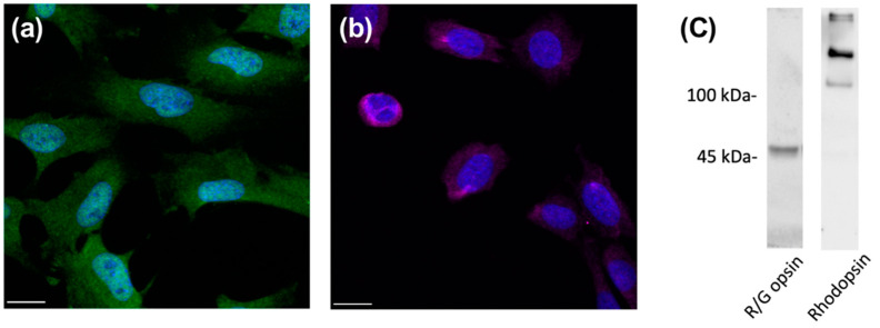
Diabetic retinopathy (DR) was identified as a leading cause of blindness and vision impairment in 2020. In addition to vasculopathy, DR has been found to involve retinal neurons, including amacrine cells and retinal ganglion cells. Despite possessing features that are susceptible to diabetic conditions, photoreceptor cells have received relatively little attention with respect to the development of DR. Until recently, studies have suggested that photoreceptors secret proinflammatory molecules and produce reactive oxygen species that contribute to the development of DR. However, the effect of hyperglycemia on photoreceptors and its underlying mechanism remains elusive. In this study, the direct effect of high glucose on photoreceptor cells was investigated using a 661w photoreceptor-like cell line. A data-independent sequential window acquisition of all theoretical mass spectra (SWATH)-based proteomic approach was employed to study changes induced by high glucose in the proteomic profile of the cells. The results indicated that high glucose induced a significant increase in apoptosis and ROS levels in the 661w cells, with mitochondrial dysfunction among the major affected canonical pathways. The involvement of mitochondrial dysfunction was further supported by increased mitochondrial fission and reduced mitochondrial bioenergetics. Collectively, these findings provide a biological basis for a possible role of photoreceptors in the pathogenesis of DR.

Keywords: SWATH-MS; diabetic retinopathy; mitochondrial dysfunction.


Conflict of interest statement

The authors declare no conflict of interest.

Figures

Figure 1
Figure 1
Expression of photoreceptor-specific markers in 661w cells. Immunocytochemical staining of 661w cells for (a) red/green opsin (green) and (b) rhodopsin (violet). DAPI (blue) was used to counterstain the nuclei. Scale bar: 20 µm. Immunoblot analysis (c) was used to confirm the expression of red/green opsin (~48 kDa) and rhodopsin (oligomeric form, >100 kDa) in 661W cells.
Figure 2
Figure 2
Effect of high-glucose apoptosis and ROS levels on the 661w cell line. Representative flow cytometry dot plots of cells incubated in (a) normal glycemic control and (b) high glucose for annexin V-PI counterstain. (c) Dot plots showing the relative change in cell apoptosis of 661w cells under the two conditions. (d) Relative change in ROS levels (CM-H2DCFDA fluorescence intensity normalized to Hoechst 33342 fluorescence intensity) of 661w cells in normal glycemic control and high glucose. (* p < 0.05, *** p < 0.001; data presented as mean ± SEM. n ≥ 9 (biological replicates)).
Figure 3
Figure 3
Volcano plot displaying the statistical p-value with the magnitude of fold change between proteins of 661w cells after 48 h incubation under high glucose vs. a normal glycemic control. The non-axial vertical dashed lines denote a fold change, of ±0.58 Log2 unit (i.e., ±1.5−fold change), whereas the non-axial horizontal dashed line denotes 1.30 −Log10 p-value (i.e., p = 0.05), which is the significance threshold prior to logarithmic transformation. A total of 992 of 2802 proteins were found to be differentially expressed (blue: downregulated; red: upregulated). The top 10 up- and downregulated proteins are labelled.
Figure 4
Figure 4
Validation of four differentially expressed proteins involved in mitochondrial dysfunction by Western blot analysis. (a) Representative Western blot (N: normal glycemic control and H: high glucose); (be) plots showing the densitometry analysis for protein levels relative to the normal glycemic control. β-actin was used as a loading control. Results illustrate the downregulation of Sod2 (~25 kDa), Snca (dimer form, ~28 kDa), and Prdx5 (~17 kDa) and the upregulation of Ogdh (~116 kDa) in 661w cells incubated in high glucose, which are consistent with our MS results. (* p < 0.05; data presented as mean ± SEM. n ≥ 4 (biological replicates) for each plot).
Figure 5
Figure 5
Effect of high glucose on mitochondrial bioenergetics of 661w cells. (a) Illustrative diagram of how key mitochondrial function parameters are divided from the Seahorse Mito Stress assay. The Seahorse XF analyzer first measures the oxygen consumption rate (OCR) as the basal respiration of the cells, and the rate of mitochondrial ATP synthesis can be estimated from the decline in OCR after adding oligomycin, an ATP synthase (complex V) inhibitor, with the remaining OCR representing the proton leak across the mitochondrial membrane in situ. The subsequent addition of an uncoupling agent, FCCP, collapses the proton gradient and disrupts the mitochondrial membrane potential, allowing the oxygen consumption of complex IV, the rate-limiting step of oxidative phosphorylation, to reach the maximum levels. This increase in OCR reflects the maximal respiration rate of the mitochondria, and the difference between the maximal respiration and basal respiration indicates the spare capacity of the mitochondria. Moreover, the injection of rotenone and antimycin A mixture ceases mitochondrial respiration, and the remaining OCR reveals the non-mitochondria-related usage of oxygen. (b) Dot plots showing the effect of high glucose on the key mitochondrial function parameters of 661w cells. (* p < 0.05. Data presented as means ± SEM. n = 5 (biological replicates)).
Figure 6
Figure 6
High-glucose-induced mitochondrial morphology changes and increased Fis1 levels and cytochrome c release in 661w cells. Representative confocal images showing mitochondrial morphology of 661w cells incubated in (a) the normal glycemic control and (b) high glucose. The scale bar represents 10 μm. Mitochondrial fragmentation was observed in 661w cells incubated with high glucose concentration. Graph showing the mean (c) form factor (FF) and (d) aspect ratio (AR) values for mitochondria of 661w cells after 48 h incubation in the two glucose concentrations. FF indicates the mitochondrial length and degree of mitochondrial branching. An FF value of one reflects a circular, unbranched mitochondrion, whereas a higher FF value implies a longer, more branched mitochondrion. AR represents the circularity of a mitochondrion, with a value of one indicating a perfect circle; the AR value increases with elongated and elliptical mitochondria. (** p < 0.01 *** p < 0.001; data presented as mean ± SEM. n ≥ 9 (biological replicates)). (e) Representative Western blot and (f) graphic illustration showing the Fis1 expression in 661w cells after normalization to the actin signal relative to the normal glycemic control. (** p < 0.01; data presented as mean ± SEM. n = 3 (biological replicates)). Cytochrome c translocation to the cytosol was assessed by Western blot analysis after subcellular fractionation. Cytosolic protein fractions were isolated from 661w cells after 48 h incubation under normal glycemic control and high-glucose conditions. (g) Representative image of Western blot analysis. (h) Graphic illustration showing the relative change in cytochrome c levels in the cytosol of 661w cells under high glucose after normalization to the actin signal (* p < 0.05; data presented as mean ± SEM. n = 3 (biological replicates) for each condition).
Figure 6
Figure 6
High-glucose-induced mitochondrial morphology changes and increased Fis1 levels and cytochrome c release in 661w cells. Representative confocal images showing mitochondrial morphology of 661w cells incubated in (a) the normal glycemic control and (b) high glucose. The scale bar represents 10 μm. Mitochondrial fragmentation was observed in 661w cells incubated with high glucose concentration. Graph showing the mean (c) form factor (FF) and (d) aspect ratio (AR) values for mitochondria of 661w cells after 48 h incubation in the two glucose concentrations. FF indicates the mitochondrial length and degree of mitochondrial branching. An FF value of one reflects a circular, unbranched mitochondrion, whereas a higher FF value implies a longer, more branched mitochondrion. AR represents the circularity of a mitochondrion, with a value of one indicating a perfect circle; the AR value increases with elongated and elliptical mitochondria. (** p < 0.01 *** p < 0.001; data presented as mean ± SEM. n ≥ 9 (biological replicates)). (e) Representative Western blot and (f) graphic illustration showing the Fis1 expression in 661w cells after normalization to the actin signal relative to the normal glycemic control. (** p < 0.01; data presented as mean ± SEM. n = 3 (biological replicates)). Cytochrome c translocation to the cytosol was assessed by Western blot analysis after subcellular fractionation. Cytosolic protein fractions were isolated from 661w cells after 48 h incubation under normal glycemic control and high-glucose conditions. (g) Representative image of Western blot analysis. (h) Graphic illustration showing the relative change in cytochrome c levels in the cytosol of 661w cells under high glucose after normalization to the actin signal (* p < 0.05; data presented as mean ± SEM. n = 3 (biological replicates) for each condition).
Figure 7
Figure 7
Schematic flowchart of the liquid chromatography-tandem mass spectrometry (LC-MS/MS) experiment of 661w cells incubated in high glucose vs. the normal glycemic control.

Similar articles

Cited by

References

    1. Klein B.E.K. Overview of epidemiologic studies of diabetic retinopathy. Ophthalmic Epidemiol. 2007;14:179–183. doi: 10.1080/09286580701396720. - DOI - PubMed
    1. Leasher J.L., Bourne R.R., Flaxman S.R., Jonas J.B., Keeffe J., Naidoo K., Pesudovs K., Price H., White R.A., Wong T.Y. Global estimates on the number of people blind or visually impaired by diabetic retinopathy: A meta-analysis from 1990 to 2010. Diabetes Care. 2016;39:1643–1649. doi: 10.2337/dc15-2171. - DOI - PubMed
    1. Barber A.J. A new view of diabetic retinopathy: A neurodegenerative disease of the eye. Prog. Neuro-Psychopharmacol. Biol. Psychiatry. 2003;27:283–290. doi: 10.1016/S0278-5846(03)00023-X. - DOI - PubMed
    1. Antonetti D.A., Barber A.J., Bronson S.K., Freeman W.M., Gardner T.W., Jefferson L.S., Kester M., Kimball S.R., Krady J.K., LaNoue K.F. Diabetic retinopathy. Diabetes. 2006;55:2401–2411. doi: 10.2337/db05-1635. - DOI - PubMed
    1. Tzekov R., Arden G. The electroretinogram in diabetic retinopathy. Surv. Ophthalmol. 1999;44:53–60. doi: 10.1016/S0039-6257(99)00063-6. - DOI - PubMed