Review
. 2022 Nov 3;23(21):13424.
doi: 10.3390/ijms232113424.

BTN3A: A Promising Immune Checkpoint for Cancer Prognosis and Treatment

Affiliations
Review

BTN3A: A Promising Immune Checkpoint for Cancer Prognosis and Treatment

Abdou-Samad Kone et al. Int J Mol Sci. .

Abstract

Butyrophilin-3A (BTN3A) subfamily members are a group of immunoglobulins present on the surface of different cell types, including innate and cancer cells. Due to their high similarity with the B7 family members, different studies have been conducted and revealed the involvement of BTN3A molecules in modulating T cell activity within the tumor microenvironment (TME). However, a great part of this research focused on γδ T cells and how BTN3A contributes to their functions. In this review, we will depict the roles and various aspects of BTN3A molecules in distinct tumor microenvironments and review how BTN3A receptors modulate diverse immune effector functions including those of CD4+ (Th1), cytotoxic CD8+ T cells, and NK cells. We will also highlight the potential of BTN3A molecules as therapeutic targets for effective immunotherapy and successful cancer control, which could represent a bright future for patient treatment.

Keywords: BTN3A; T lymphocytes; cancer; immune system; prognostic factor.


Conflict of interest statement

The authors declare no conflict of interest.

Figures
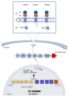
Figure 1
Figure 1
Schematic representation of the CD277/BTN3A gene, mRNA, and protein structural domains. (A) The extracellular domain of interaction with the ligand. (B) The transmembrane domain. (C) The intracellular domain. BTN3A1 and BTN3A3 share the B30.2 domain involved in the interaction with the phosphoantigens.
Figure 2
Figure 2
BTN3A interaction patterns and signaling pathways. BTN3A-dependent co-stimulatory mechanisms: (1) engagement of BTN3A with its putative ligand on APC and/or (2) treatment of BTN3A with CD277 mAb 20.1 provides a co-stimulatory T cell activation signal that induces production of IL-2 and IFNγ as well as T cell proliferation via the PI3K/AKT signaling pathway. BTN3A-dependent co-inhibitory mechanisms; (3) interaction of BTN3A with a specific mAb 232.5 suppresses c-FLIP expression. The cellular inhibitory protein type FLICE-like (c-FLIP) is well-known to promote the maturation and survival of T cells through interaction with pro-caspase, which activates the activation cascade ERK/NF-κB. The inhibition of c-FLIP expression by BTN3A triggers an inhibiting signal that interferes with the production of IFNγ, IL-4, and the proliferation of T cells. (4) BTN3A1 binds the N-mannosylated residues of CD45 and, thus, elicits the inhibition of the TCR signal on T cells. CD45 is well recognized to play a key role in T cell activation through the TCR signaling pathway. The BTN3A1 and BTN3A2 provide differential regulation of NKp30-induced IFNγ production: (5) The co-engagement of BTN3A1 with NKp30 after treatment with anti-NKp30 mAb modulates the NKp30-induced IFN-γ production, (6) whereas the lack of B30.2 domain in the BTN3A2 structure impair the co-engagement of BTN3A2 with NKp30 and thus lead to the attenuation of the NKp30-induced IFN-γ production. LSECtin on tumor-associated macrophages improves the stemness of tumor cells through interaction with BTN3A receptor: (7) BTN3A1/2 Interaction with LSECtin in tumor cells actives the JAK2/STAT3 signaling pathway, which promotes cancer cell survival, invasion, and proliferation.

References

    1. Liu M., Guo F. Recent updates on cancer immunotherapy. Precis. Clin. Med. 2018;1:65–74. doi: 10.1093/pcmedi/pby011. - DOI - PMC - PubMed
    1. Wherry E.J. T cell exhaustion. Nat. Immunol. 2011;12:492–499. doi: 10.1038/ni.2035. - DOI - PubMed
    1. Jiang W., He Y., He W., Wu G., Zhou X., Sheng Q., Zhong W., Lu Y., Ding Y., Lu Q., et al. Exhausted CD8+T Cells in the Tumor Immune Microenvironment: New Pathways to Therapy. Front. Immunol. 2020;11:622509. doi: 10.3389/fimmu.2020.622509. - DOI - PMC - PubMed
    1. Borghaei H., Paz-Ares L., Horn L., Spigel D.R., Steins M., Ready N.E., Chow L.Q., Vokes E.E., Felip E., Holgado E., et al. Nivolumab versus Docetaxel in Advanced Nonsquamous Non-Small-Cell Lung Cancer. N. Engl. J. Med. 2015;373:1627–1639. doi: 10.1056/NEJMoa1507643. - DOI - PMC - PubMed
    1. Seidel J.A., Otsuka A., Kabashima K. Anti-PD-1 and Anti-CTLA-4 Therapies in Cancer: Mechanisms of Action, Efficacy, and Limitations. Front. Oncol. 2018;8:86. doi: 10.3389/fonc.2018.00086. - DOI - PMC - PubMed