. 2022 Nov 3;23(21):13458.
doi: 10.3390/ijms232113458.

Proteomics of Aqueous Humor as a Source of Disease Biomarkers in Retinoblastoma

Affiliations

Proteomics of Aqueous Humor as a Source of Disease Biomarkers in Retinoblastoma

Angela Galardi et al. Int J Mol Sci. .

Abstract

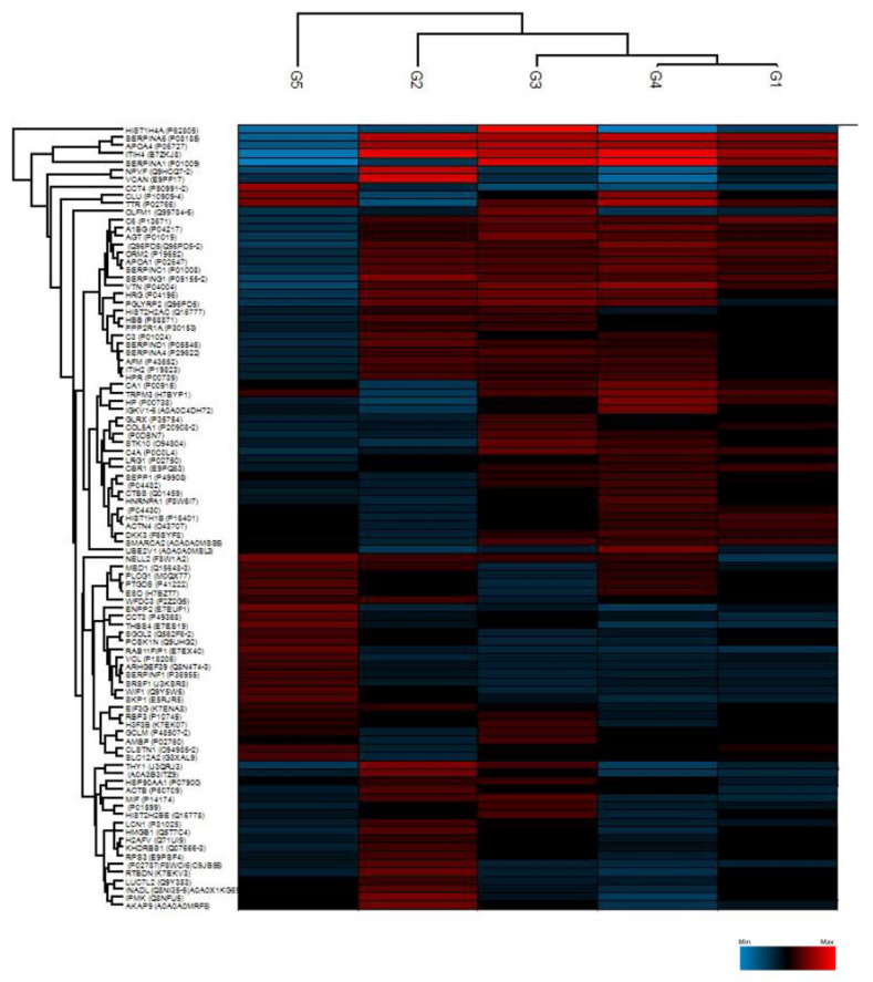
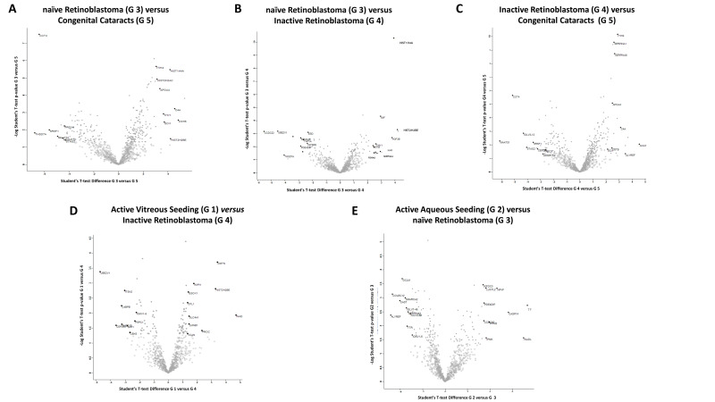
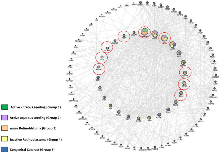
Aqueous humor (AH) can be easily and safely used to evaluate disease-specific biomarkers in ocular disease. The aim of this study was to identify specific proteins biomarkers in the AH of retinoblastoma (RB) patients at various stages of the disease. We analyzed the proteome of 53 AH samples using high-resolution mass spectrometry. We grouped the samples according to active vitreous seeding (Group 1), active aqueous seeding (Group 2), naive RB (group 3), inactive RB (group 4), and congenital cataracts as the control (Group 5). We found a total of 889 proteins in all samples. Comparative parametric analyses among the different groups revealed three additional proteins expressed in the RB groups that were not expressed in the control group. These were histone H2B type 2-E (HISTH2B2E), InaD-like protein (PATJ), and ubiquitin conjugating enzyme E2 V1 (UBE2V1). Upon processing the data of our study with the OpenTarget Tool software, we found that glyceraldehyde 3-phosphate dehydrogenase (GAPDH) and CD44 were more highly expressed in the RB groups. Our results provide a proteome database regarding AH related to RB disease that may be used as a source of biomarkers. Further prospective studies should validate our finding in a large cohort of RB patients.

Keywords: aqueous humor; proteomic; retinoblastoma; tumor biomarker.


Conflict of interest statement

The authors declare that they have no conflict of interest.

Figures

Figure 1
Figure 1
Unsupervised hierarchical-clustered heatmap of proteins identified by ANOVA testing. Heatmap showing the distinctive proteomic signature of the 5 different groups: Group 1 (active vitreous seeding), Group 2 (active aqueous seeding), Group 3 (naïve RB), Group 4 (inactive RB), and Group 5 (congenital cataracts). The amount of each protein in the different groups is represented by the color scheme, in which red and blue indicate high and low proteins expression, respectively.
Figure 2
Figure 2
Volcano plots showing abundance of differentially expressed proteins between two of the five groups at a time: (A) naïve RB compared to congenital cataracts; (B) naïve RB compared to inactive RB; (C) inactive RB compared to congenital cataracts; (D) active vitreous seeding compared to inactive RB; (E) active aqueous seeding compared to naïve RB. The black line shows p value = 0.05 and s0 = 0.1. Red dots depict proteins whose fold change is <2 (log2 = 1) or p > 0.05 between the two groups considered.
Figure 3
Figure 3
The network represents the interactions between 96 genes coding proteins identified overlapping the polypeptides associated with RB, selected via Open Target, and the proteins identified in our study groups (including the congenital cataract group). The nodes have dimensions related to the degree of connection, and the internal circumference shows the most connected proteins, with a degree > 15. The outer circumference shows the less connected proteins (degree ≤ 15); therefore, of smaller dimensions. Connections are only present if the confidence level of the string is > 0.4. The coloring of the nodes is a function of the average intensity of each group measured in mass spectrometry, normalized for graphic reasons on a scale of 1–100. The coloring and the percentage distribution of the node were preformed using the layout tools in Image/Chart of Cytoscape.
Figure 4
Figure 4
Workflow of the proteomic analysis of the aqueous humor in patients affected by retinoblastoma. The figure was created with https://biorender.com.

References

    1. Kivelä T. The epidemiological challenge of the most frequent eye cancer: Retinoblastoma, an issue of birth and death. Br. J. Ophthalmol. 2009;9:1129–1131. doi: 10.1136/bjo.2008.150292. - DOI - PubMed
    1. Dyson N.J. RB1: A prototype tumor suppressor and an enigma. Genes Dev. 2016;30:1492–1502. doi: 10.1101/gad.282145.116. - DOI - PMC - PubMed
    1. Munier F.L., Soliman S., Moulin A.P., Gaillard M.C., Balmer A., Beck-Popovic M. Profiling safety of intravitreal injections for retinoblastoma using an anti-reflux procedure and sterilisation of the needle track. Br. J. Ophthalmol. 2012;8:1084–1087. doi: 10.1136/bjophthalmol-2011-301016. - DOI - PubMed
    1. Berry J.L., Xu L., Murphree A.L., Krishnan S., Stachelek K., Zolfaghari E., McGovern K., Lee T.C., Carlsson A., Kuhn P., et al. Potential of Aqueous Humor as a Surrogate Tumor Biopsy for Retinoblastoma. JAMA Ophthalmol. 2017;135:1221–1230. doi: 10.1001/jamaophthalmol.2017.4097. - DOI - PMC - PubMed
    1. Raval V., Racher H., Wrenn J., Singh A.D. Aqueous humor as a surrogate biomarker for retinoblastoma tumor tissue. J. AAPOS. 2022;13:137.e1–137.e5. doi: 10.1016/j.jaapos.2022.03.005. - DOI - PubMed