. 2022 Nov 3;23(21):13464.
doi: 10.3390/ijms232113464.

Physiological Impact of a Synthetic Elastic Protein in Arterial Diseases Related to Alterations of Elastic Fibers: Effect on the Aorta of Elastin-Haploinsufficient Male and Female Mice

Affiliations

Physiological Impact of a Synthetic Elastic Protein in Arterial Diseases Related to Alterations of Elastic Fibers: Effect on the Aorta of Elastin-Haploinsufficient Male and Female Mice

Quentin Boëté et al. Int J Mol Sci. .

Abstract

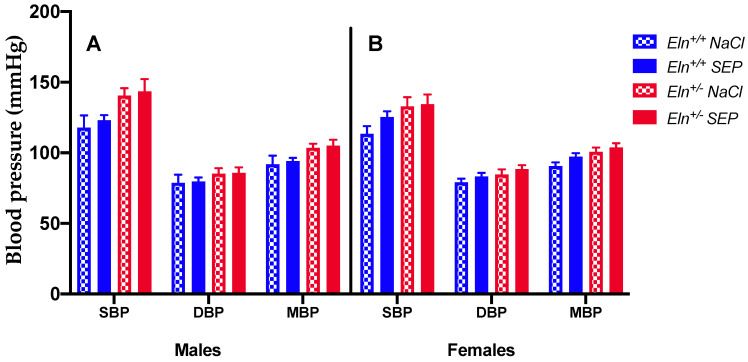
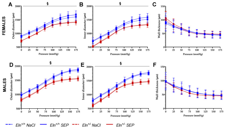
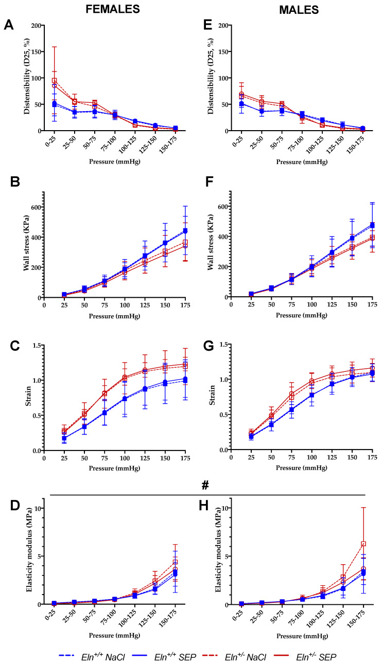
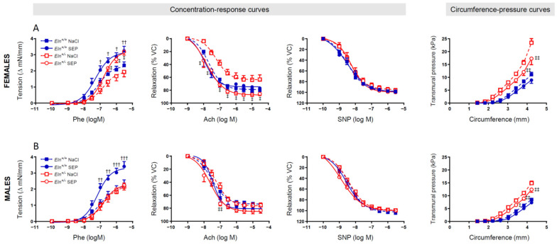
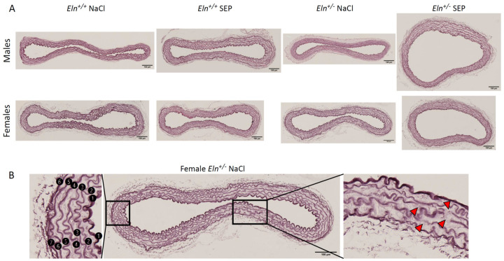
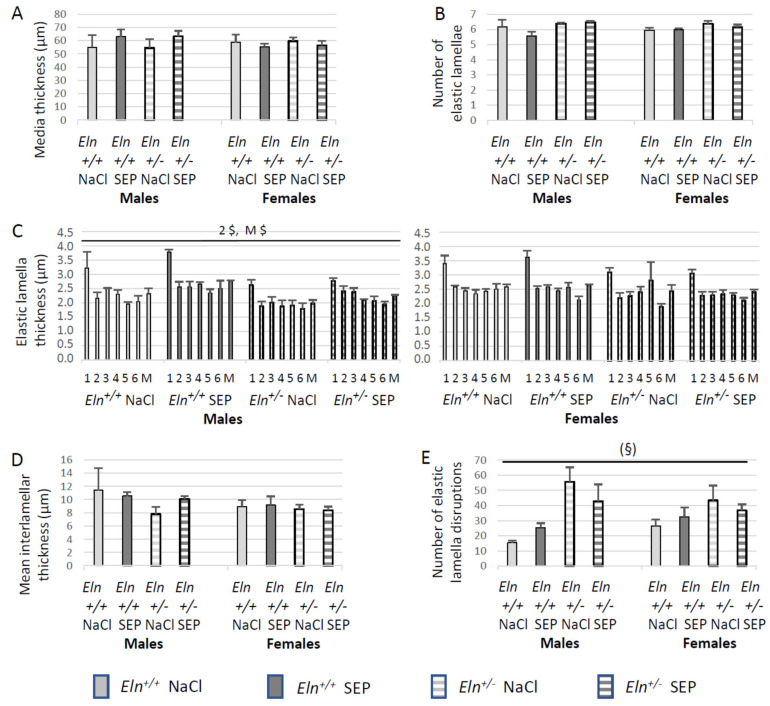
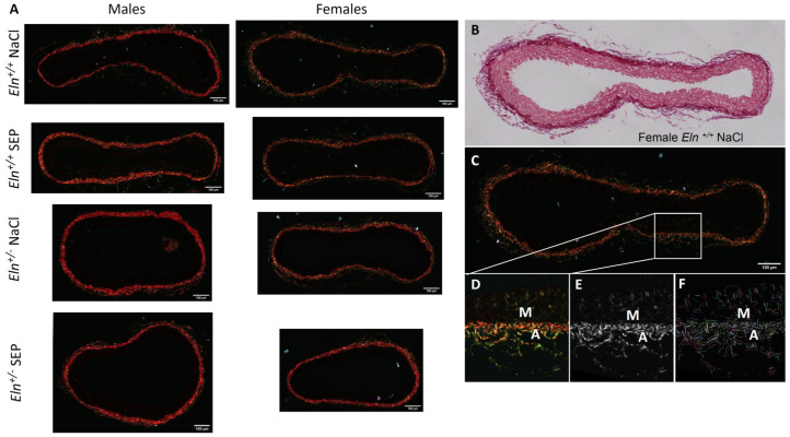
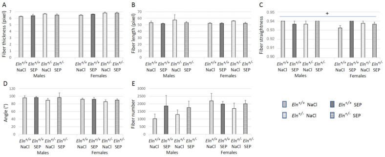
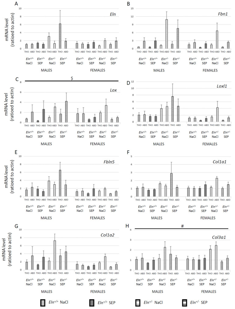
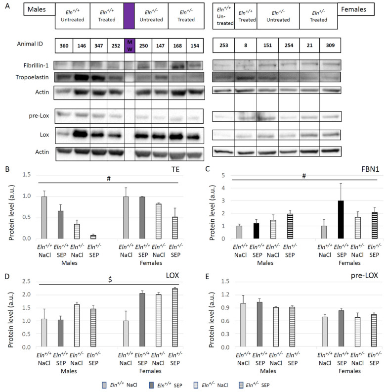
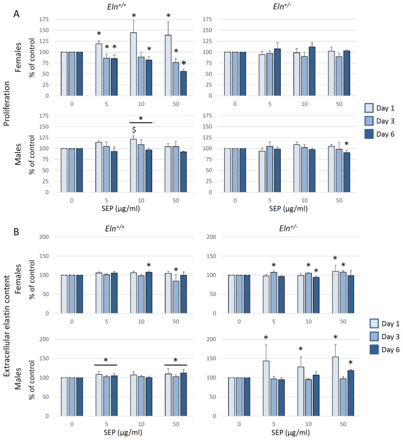
Elastic fibers, made of elastin (90%) and fibrillin-rich microfibrils (10%), are the key extracellular components, which endow the arteries with elasticity. The alteration of elastic fibers leads to cardiovascular dysfunctions, as observed in elastin haploinsufficiency in mice (Eln+/-) or humans (supravalvular aortic stenosis or Williams-Beuren syndrome). In Eln+/+ and Eln+/- mice, we evaluated (arteriography, histology, qPCR, Western blots and cell cultures) the beneficial impact of treatment with a synthetic elastic protein (SEP), mimicking several domains of tropoelastin, the precursor of elastin, including hydrophobic elasticity-related domains and binding sites for elastin receptors. In the aorta or cultured aortic smooth muscle cells from these animals, SEP treatment induced a synthesis of elastin and fibrillin-1, a thickening of the aortic elastic lamellae, a decrease in wall stiffness and/or a strong trend toward a reduction in the elastic lamella disruptions in Eln+/- mice. SEP also modified collagen conformation and transcript expressions, enhanced the aorta constrictive response to phenylephrine in several animal groups, and, in female Eln+/- mice, it restored the normal vasodilatory response to acetylcholine. SEP should now be considered as a biomimetic molecule with an interesting potential for future treatments of elastin-deficient patients with altered arterial structure/function.

Keywords: aorta; elastic fiber synthesis/repair; elastin haploinsufficiency; mechanics; pharmacotherapy; reactivity; structure; synthetic elastic protein.


Conflict of interest statement

The authors declare no conflict of interest. The funders had no role in the design of the study; the collection, analyses, or interpretation of data; the writing of the manuscript, or in the decision to publish the results.

Figures

Figure 1
Figure 1
Systolic, diastolic and mean arterial blood pressures. The measurements were performed 3 days after NaCl (control) or SEP intravenous injection in the caudal vein of male and female mice of Eln+/+ or Eln+/- genotypes. (A) Males, (B) Females, SBP: Systolic Blood Pressure, DBP: Diastolic Blood Pressure, MBP: Mean Blood Pressure. No significant effect of SEP treatment could be detected (four-way ANOVA and three-way ANOVAs for each sex, p > 0.05). Control animals were injected with the same volume of 0.9% NaCl. Values are mean ± SEM. n = 4–7 per group.
Figure 2
Figure 2
Diameter–pressure and wall-thickness–pressure curves of the cannulated ascending aorta. The measurements were performed by pressure arteriography 3 days after NaCl (control) or SEP intravenous injection in the caudal vein of male and female mice of Eln+/+ or Eln+/- genotypes ((AC): females; (DF): males). (A,D): outer-diameter–pressure relation. (B,E): inner-diameter–pressure relation. (C,F): wall-thickness–pressure relation. § general significant interaction between genotype and treatment (three-way ANOVA, p ≤ 0.05), showing that SEP treatment induced a diameter elevation in Eln+/+, not Eln+/-, mice, independently of sex (post hoc Fisher’s least significant difference (LSD) tests, p ≤ 0.05). Control animals were injected with the same volume of 0.9% NaCl. Values are mean ± SEM. n = 7–10 per group.
Figure 3
Figure 3
Mechanical parameters of the cannulated ascending aorta. The measurements were performed by pressure arteriography 3 days after NaCl (control) or SEP intravenous injection in the caudal vein of male and female mice of Eln+/+ or Eln+/- genotypes. (A,E) Aortic distensibility (D25)–pressure increment relation; (B,F) circumferential stress–pressure relation; (C,G) circumferential strain–pressure relation; (D,H) incremental elastic modulus (Einc)–pressure increment relation. # general significant effect of treatment (NaCl control vs. SEP), independently of genotype, sex and pressure (four-way ANOVA, p ≤ 0.05). Control animals were injected with the same volume of 0.9% NaCl. Values are mean ± SEM. n = 7–10 per group.
Figure 4
Figure 4
Concentration–response and circumference–pressure curves of the ascending aorta. The studies were performed by wire myography in aortae from female (A) and male (B) Eln+/+ and Eln+/- mice treated with a single 0.9% NaCl (controls) or synthetic elastic protein (SEP) intravenous injection three days before the measurements.  p < 0.05, †† p < 0.01 and ††† p < 0.001: significant differences between Eln+/+SEP and Eln+/+NaCl;  p < 0.05 and ‡‡ p < 0.01: significant differences between Eln+/-SEP and Eln+/-NaCl (two-way ANOVAs, followed, where necessary, by multiple Bonferroni post-tests). Values are mean ± SEM. n = 11–14 in each NaCl group; n = 7–8 in each SEP group.
Figure 5
Figure 5
Elastic lamella detection by orcein staining in cross-sections of the distal part of the aortic arch of Eln+/+ or Eln+/- male or female mice injected with SEP or 0.9% NaCl (control). (A) Representative images for each group. (B) Descriptive example, for an Eln+/- female mouse injected with 0.9% NaCl (control), of the process used for the quantification of elastic lamella abnormalities: (i) on the left, the numbering of each elastic lamella from the luminal side (#1: Internal elastic lamina) to the external side of the media (#6 or #7, depending on the vessel), and (ii) on the right, the identification of elastic lamella disruptions (red arrowheads). Scale bars = 100 µm.
Figure 6
Figure 6
Analysis of the morphology and structure of the orcein-stained elastic fibers/elastic lamellae present in the distal part of the aortic arch, depending on genotype, sex and SEP treatment. The measurements were performed three days after NaCl (control) or SEP intravenous injection in the caudal vein of male and female mice of Eln+/+ or Eln+/- genotypes. (A) Media thickness. (B) Number of elastic lamellae. (C) Mean thickness of the elastic lamellae (M) and thickness of each elastic lamellae, numbered from the luminal side of the media to the external side of the media, lamella #1 being the internal elastic lamina (see Figure 5 for illustration). (D) Mean of interlamellar thickness. (E) Number of elastic lamella disruptions. $ significant interaction between sex and treatment (three-way ANOVA, p ≤ 0.05), showing that SEP treatment induced an increase in elastic lamella #2 thickness (2 $) or mean elastic lamella thickness (M $) in males, not females, independently of genotype (post hoc Fisher’s least significant difference (LSD) tests, p ≤ 0.05). (§) strong trend toward an interaction between genotype and SEP treatment, independently of sex (three-way ANOVA, p < 0.08), suggesting a lower number of elastic lamella disruptions in Eln+/- mice after SEP treatment. Control animals were injected with the same volume of 0.9% NaCl (NaCl). Values are mean ± SEM. n = 3–6 in each group.
Figure 7
Figure 7
Images of picrosirius red-stained collagen fibers in cross-sections of the distal part of the aortic arch of Eln+/+ or Eln+/- male or female mice injected with SEP or 0.9% NaCl (control), illuminated with polarized light (A). Representative example of an aorta from an Eln+/+ female mouse injected with 0.9% NaCl (BF), under illumination with non-polarized light (B), compared to polarized light (CF). A magnified image of the selected area of the aorta wall shown in (C) is presented in (D). The images from the whole aorta wall were then CT-FIRE-processed in order to obtain the individual fiber properties and quantity of the collagen fibers in all the studied groups (fiber width, length, straightness, angle and number of fibers in the whole aorta section). The second harmonic generation (SHG) image generated from (D) using CT-FIRE program is shown in (E). The extracted fibers obtained using the CT-FIRE program are highlighted in different colors overlaid on the original SHG image in F. M = Media, A = Adventitia. Scale bars = 100 µm.
Figure 8
Figure 8
CT-FIRE analysis of picrosirius red-stained cross-sections of the distal part of the aortic arch (entire aorta wall section) illuminated with polarized light. The measurements were performed 3 days after NaCl (control) or SEP intravenous injection in the caudal vein of male and female mice of Eln+/+ or Eln+/- genotypes. Individual properties and quantity of collagen fibers were compared between the studied groups: (A) fiber thickness, (B) length, (C) straightness, (D) angle and (E) number of fibers per aorta. + significant interaction between sex, genotype and SEP treatment (three-way ANOVA, p ≤ 0.05), showing that the elevation of collagen fiber straightness in untreated Eln+/+ males, compared to Eln+/+ female mice, was abolished by the SEP-induced increase in collagen fiber straightness in female Eln+/+ mice (post hoc Fisher’s least significant difference (LSD) tests, p ≤ 0.05). Control animals were injected with the same volume of 0.9% NaCl (NaCl). Values are mean ± SEM. n = 3–6 in each group.
Figure 9
Figure 9
Effect of SEP treatment on the levels of mRNAs transcribed from the genes of the major components of aortic elastic and collagen fibers. The measurements were performed by RT-qPCR in thoracic (THO) and abdominal (ABD) aorta segments 3 days after SEP or NaCl (control) intravenous injection in the caudal vein of male and female mice of Eln+/+ or Eln+/- genotype. (A) tropoelastin (Eln), (B) fibrillin-1 (Fbn1), (C) lysyl oxidase (Lox), (D) lysyl oxidase-like-1 (Loxl1), (E) fibulin-5 (Fbln5), (F) type I collagen alpha-1 (Col1a1), (G) type I collagen alpha-2 (Col1a2), (H) type III collagen alpha-1 (Col3a1). $ significant interaction between sex and SEP treatment, independently of genotype and vessel segment (four-way ANOVA, p ≤ 0.05), indicating that SEP significantly decreased mRNA levels in female, not male, mice (post hoc Fisher’s least significant difference (LSD) tests, p ≤ 0.05). # general significant difference between untreated and SEP-treated mice, independently of sex, genotype and vessel segment (four-way ANOVA, p ≤ 0.05). Control animals were injected with the same volume of 0.9% NaCl (NaCl). Values are mean ± SEM. n = 5 in each group.
Figure 10
Figure 10
Western blotting of descending thoracic aorta extracts from untreated or SEP-treated mice. (A) Representative Western blot bands for the proteins of interest in each group. Semi-quantitative analysis of the protein levels was performed by band densitometry for: (B) tropoelastin (TE), (C) fibrillin-1 (FBN1), (D) lysyl oxidase (LOX, 30kDa), (E) immature lysyl oxidase (pre-LOX, 50kDa). # general significant difference between untreated and SEP-treated mice (three-way ANOVA, p ≤ 0.05). $ significant interaction between sex and treatment, independently of genotype (three-way ANOVA, p ≤ 0.05), indicating that SEP elevated the protein level in females, not males (post hoc Fisher’s least significant difference (LSD) tests, p ≤ 0.05). Control animals were injected with the same volume of 0.9% NaCl (NaCl). Values are mean ± SEM. n = 5 in each group.
Figure 11
Figure 11
Proliferation and extracellular elastin content in primary cultures of aortic VSMCs from male or female Eln+/+ or Eln+/- mice. VSMCs were treated with SEP (0, 5, 10 or 50 µg/mL) for 1, 3 or 6 days. (A) Impact of SEP treatment on VSMC proliferation, evaluated by using the MTT assay. The results are expressed as percent of the control value (0 µg/mL SEP) of the same day. (B) Impact of SEP treatment on extracellular elastin content, evaluated by using an ELISA assay. The results are expressed as percent of the control value (0 mg/mL SEP) of the same day. * significant difference with same day control (three-way ANOVA, followed by LSD test for paired comparisons, p ≤ 0.05). $ significant difference at day 1 with same day control (two-way ANOVA, followed by LSD test for paired comparisons, p ≤ 0.05). Values are mean ± SEM. n = 3–5 primary cultures from separate animals in each group.

References

    1. Tucker W.D., Mahajan K. StatPearls. StatPearls Publishing; Treasure Island, FL, USA: 2019. Anatomy, blood vessels. - PubMed
    1. Greenwald S.E. Ageing of the Conduit Arteries. J. Pathol. 2007;211:157–172. doi: 10.1002/path.2101. - DOI - PubMed
    1. O’Rourke M.F., Hashimoto J. Mechanical Factors in Arterial Aging: A Clinical Perspective. J. Am. Coll. Cardiol. 2007;50:1–13. doi: 10.1016/j.jacc.2006.12.050. - DOI - PubMed
    1. Chapter 7: The Cardiovascular System: Blood Vessels and Circulation—Anatomy & Physiology. [(accessed on 18 May 2022)]. Available online: https://qut.pressbooks.pub/anatomyandphysiology/chapter/chapter-20-the-c...
    1. Behmoaras J., Osborne-Pellegrin M., Gauguier D., Jacob M.-P. Characteristics of the Aortic Elastic Network and Related Phenotypes in Seven Inbred Rat Strains. Am. J. Physiol. Heart Circ. Physiol. 2005;288:H769–H777. doi: 10.1152/ajpheart.00544.2004. - DOI - PubMed